Ameloblastoma: Jaw Tumour Symptoms, Diagnosis & Surgery
Need expert consultation? Book an appointment with Dr. Pradeep S. or Dr. Kalpa Pandya.
Book AppointmentDiscovering a lump, swelling, or unusual pain in your jaw can be deeply unsettling. While many jaw swellings are related to common dental infections, some arise from deeper, structural tissues. One such condition is ameloblastoma, a rare and complex jaw tumour.
Understanding the clinical pathway of ameloblastoma jaw tumour symptoms diagnosis and surgery is the first step toward successful treatment and recovery. Because these tumours are locally aggressive, early identification and precise surgical planning are crucial to preserving your facial structure, speech, and ability to eat.
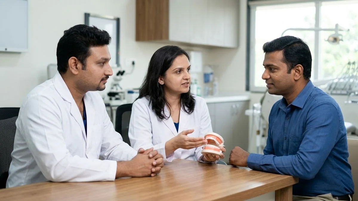
At Mouth Cancer Surgeons in Chennai, our dual-surgeon team—Dr. Pradeep S. and Dr. Kalpa Pandya—works collaboratively to guide patients through every phase of care, from initial diagnostic imaging to advanced microvascular jaw reconstruction.
What is Ameloblastoma? Understanding This Jaw Tumour
An ameloblastoma is a rare, non-cancerous (benign) tumour that develops primarily in the jawbone. It originates from ameloblasts, the specialized cells responsible for producing tooth enamel during dental development. Although it is classified as benign because it rarely spreads to distant organs, it behaves aggressively within the local facial skeleton.
Ameloblastomas are osteolytic, meaning they actively break down and destroy the surrounding bone as they grow. If left untreated, the tumour can expand significantly, causing severe facial deformity, destroying the jaw joint, moving teeth out of alignment, and invading adjacent structures like the nasal cavity, maxillary sinuses, or orbit.
These tumours most commonly occur in the lower jaw (mandible), particularly near the region of the lower third molars (wisdom teeth) and the ascending ramus of the jaw. However, they can also develop in the upper jaw (maxilla), where they tend to be even more aggressive due to the thin, porous nature of the maxillary bones. Ameloblastomas can affect individuals of any age, though they are most frequently diagnosed in adults between their third and fifth decades of life.
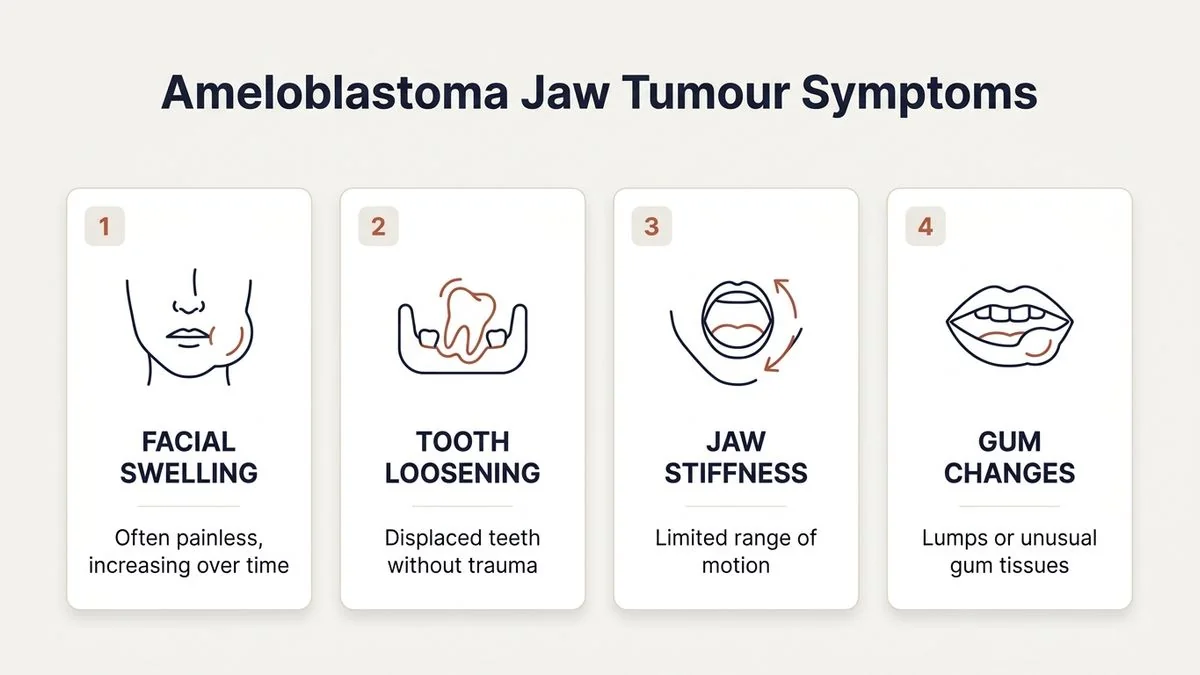
Spotting the Warning Signs: Ameloblastoma Jaw Tumour Symptoms
In its earliest stages, an ameloblastoma is often completely silent. Many patients only discover the tumour incidentally during routine dental check-ups, when a panoramic X-ray (OPG) reveals an unexpected dark shadow inside the jawbone.
As the tumour slowly expands and thins the overlying bone, distinct physical symptoms begin to emerge.
Early-Stage Symptoms
- Painless Jaw Swelling: The most characteristic sign is a slow-growing, painless expansion of the jawbone, usually felt inside the mouth or along the lower border of the jaw or cheek.
- Tooth Mobility: Teeth in the immediate vicinity of the tumour may gradually become loose, tilt, or drift out of their normal positions without any history of gum disease.
- Delayed Tooth Eruption: In younger patients, the tumour may block a permanent tooth (often a lower wisdom tooth) from erupting naturally.
Advanced-Stage Symptoms
- Facial Asymmetry: Significant swelling can alter the natural contours of the face, causing one side of the jaw or cheek to appear visibly larger or distorted.
- Malocclusion: As the jawbone deforms and teeth shift, your upper and lower teeth may no longer fit together properly when you bite down.
- Dull Ache or Pressure: While pain is rarely the first symptom, advanced tumours can cause a persistent, dull ache or a sensation of heavy pressure within the face.
- Numbness (Paresthesia): If the growing tumour compresses the inferior alveolar nerve—the nerve running inside the lower jaw—you may experience numbness, tingling, or a "pins-and-needles" sensation in your lower lip and chin.
- Difficulty Chewing or Swallowing: Large tumours can restrict the movement of the jaw, making it difficult to chew, speak, or swallow comfortably.
If you are experiencing any unexplained jaw swelling or shifting teeth, early evaluation is essential. Request a comprehensive consultation with Dr. Pradeep S. and Dr. Kalpa Pandya at Apollo Main Hospital, Greams Road, Chennai.
How is Ameloblastoma Diagnosed? The Clinical Pathway
Because ameloblastomas can closely mimic other benign jaw cysts (such as dentigerous cysts or odontogenic keratocysts) on standard X-rays, a structured, multi-step diagnostic protocol is required to establish an accurate diagnosis.
1. Clinical Examination
Your surgeon will carefully palpate your jaw, check for areas of bone thinning (which sometimes feel like eggshell crackling under pressure), evaluate the stability of your teeth, and assess the function of your facial nerves.
2. Advanced Radiographic Imaging
- Orthopantomogram (OPG): A panoramic X-ray that provides a broad view of the entire upper and lower jaw. Ameloblastomas typically appear as distinct, multi-chambered lucencies, often described as having a "soap bubble" or "honeycomb" appearance.
- Computed Tomography (CT) Scan: A high-resolution 3D CT scan is indispensable. It maps the precise boundaries of the tumour, shows how much of the jawbone has been destroyed, and identifies any breaches in the outer cortical bone. This scan serves as the blueprint for surgical planning.
- Magnetic Resonance Imaging (MRI): If the tumour is located in the upper jaw (maxilla) or shows signs of invading nearby soft tissues, an MRI may be ordered to assess soft tissue involvement.
3. Tissue Biopsy
A biopsy is the only definitive way to confirm an ameloblastoma. Under local anaesthesia, your surgeon will make a small incision to access the jawbone, create a tiny window in the bone, and remove a small sample of the tumour tissue. This sample is sent to an oral pathologist, who examines it under a microscope to identify the specific cellular patterns of the tumour.
Types of Ameloblastoma and Why They Matter
Ameloblastomas are classified into different clinical and histological types. Identifying the specific type is critical because it directly dictates how aggressively your surgical team must treat the lesion.
| Ameloblastoma Type | Growth Characteristics | Local Aggressiveness | Recurrence Risk | Primary Surgical Approach |
|---|---|---|---|---|
| Unicystic | Consists of a single cyst-like cavity; often associated with an impacted tooth. | Moderate | Low to Moderate | Conservative (enucleation) or localized resection depending on the cyst wall involvement. |
| Multicystic / Solid | Multiple interconnected bone cavities filled with tumour tissue; most common type. | High | High (if not widely resected) | Radical resection with 1 to 1.5 cm healthy bone margins. |
| Peripheral | Occurs entirely in the soft tissue of the gums; does not invade the underlying bone. | Low | Low | Simple local excision with a narrow margin of surrounding tissue. |
Surgical Treatment Options for Ameloblastoma
Surgery is the primary and most effective treatment for ameloblastoma. Because these tumours do not respond predictably to radiation therapy or chemotherapy, complete surgical removal is necessary.
The surgical strategy is selected based on the tumour's location, size, and specific histological type.
Conservative Surgery (Enucleation and Curettage)
For certain small, unicystic ameloblastomas, a conservative approach may be considered. This involves scooping out the tumour (enucleation) and scraping the bony cavity clean (curettage). While this preserves the jaw's structure, it carries a high risk of recurrence if even a single microscopic tumour cell is left behind.
To lower this risk, chemical agents like Carnoy's solution or physical agents like liquid nitrogen (cryotherapy) may be applied to the bone cavity to destroy any remaining microscopic cells.
Radical Resection
For solid or multicystic ameloblastomas, radical surgical resection is the gold standard. To ensure the tumour does not return, the surgeon removes the affected segment of the jawbone along with a safety margin of healthy bone (usually 1 to 1.5 centimetres beyond the visible margins of the tumour).
- Mandibulectomy: Removal of a portion of the lower jawbone. Learn more about the surgical steps involved in a mandibulectomy.
- Maxillectomy: Removal of a portion of the upper jawbone.
While radical resection is highly effective at preventing recurrence, it leaves a structural gap in the jawbone that requires immediate or staged reconstruction to restore the patient's appearance and oral function.
Jaw Reconstruction and Restorative Surgery
Modern oral and maxillofacial surgery does not just focus on removing the tumour; it places equal emphasis on rebuilding what was lost. Today, reconstructive surgery is often performed during the same operation as the tumour removal, ensuring the patient wakes up with an intact jaw.
Microvascular Free Flap Reconstruction
The gold standard for rebuilding a resected jaw is microvascular free tissue transfer. The most common donor site is the fibula (the thin bone on the outer side of the lower leg).
- Harvesting: A segment of the fibula bone, along with its blood vessels (artery and vein) and overlying skin, is carefully harvested.
- Shaping: The straight fibula bone is precisely shaped and bent to match the natural curve of the missing jaw segment.
- Anastomosis: Under an operating microscope, the surgeon connects the blood vessels of the leg graft to blood vessels in the neck. This immediately restores blood flow to the newly reconstructed jawbone, allowing it to heal and integrate like a natural jaw.
You can read more about how these procedures are planned on our dedicated reconstructive & restorative surgery page.
Dental Rehabilitation
Once the reconstructed bone has fully integrated and healed (typically 4 to 6 months after surgery), dental implants can be surgically placed directly into the reconstructed bone. These implants serve as solid anchors for custom-made crowns or bridges, fully restoring the patient's ability to bite, chew, and speak naturally.
To understand how dental implants are integrated into post-tumour care, visit our guide on dental implants & pre-prosthetic surgery.
Recovery, Prognosis, and Long-Term Surveillance
Recovering from ameloblastoma surgery is a gradual process that requires patience and a structured rehabilitation plan.
Immediate Post-Operative Phase
If you undergo a segmental resection with microvascular reconstruction, you will spend the first few days in the hospital's high-dependency unit. Your surgical team will closely monitor the blood flow to the reconstructed jaw graft. You will receive intravenous fluids, pain management medications, and nutrition through a temporary feeding tube to allow the surgical sites in your mouth to heal without irritation.
Transitioning to Home Care
Most patients are discharged within 7 to 10 days. During the first few weeks at home, you will need to follow a strict liquid or soft-food diet to avoid placing stress on the healing bone. Gentle oral hygiene practices, such as using warm saline rinses and a soft-bristled toothbrush, are crucial to prevent infection.
The Importance of Long-Term Follow-Up
Even with radical resection and clear surgical margins, ameloblastomas can occasionally recur, sometimes up to 10 or 15 years after the initial surgery. Because of this slow-growing nature, long-term surveillance is absolutely essential.
We recommend a structured follow-up schedule:
- Clinical exams and imaging (X-rays or CT scans) every 6 months for the first 2 to 3 years.
- Annual imaging studies for up to 10 years post-surgery.
Our dual-surgeon model ensures that the same two specialists who performed your surgery are there to manage your long-term follow-up visits, providing consistent, highly personalized surveillance. Learn more about our comprehensive approach to multidisciplinary oncology care & surveillance.
Choosing the Right Maxillofacial Surgeons in Chennai
Treating an ameloblastoma requires a highly specialized skill set that sits at the intersection of head and neck oncology, oral and maxillofacial surgery, and reconstructive microvascular surgery.
At Mouth Cancer Surgeons, Chennai, we offer a unique dual-surgeon care model. When you choose our practice, you are not cared for by a rotating team of junior doctors. Instead, the same two dedicated specialists manage your entire journey:
- Dr. Pradeep S. is an international board-certified Oral & Maxillofacial Surgeon with over 7 years of specialized focus in head and neck surgical oncology. He leads our complex jaw resections and reconstructive procedures.
- Dr. Kalpa Pandya brings over 10 years of experience in managing oral potentially malignant disorders, maxillofacial trauma, and dental implant rehabilitation, ensuring your post-surgical dental restoration is seamless.
Our primary surgical consultations and procedures are hosted at Apollo Main Hospital, Greams Road, Chennai, a world-class facility equipped with advanced intensive care units, state-of-the-art operating theatres, and 3D virtual surgical planning software that allows us to map out your reconstruction with sub-millimeter precision before we even enter the operating room.
For personalized treatment options and expert care, consult Dr. Pradeep S. and Dr. Kalpa Pandya — Mouth Cancer Surgeons, Chennai. Call +91 96633 03747 or book an appointment online today.
References
- McClary, Amanda C., et al. "Ameloblastoma: a clinical review and trends in management." European Archives of Oto-Rhino-Laryngology, 2016. https://link.springer.com/article/10.1007/s00405-015-3631-8
- Kreppel, M., et al. "Ameloblastoma of the jaws: an analysis of treatment outcomes and recurrence factors." Journal of Cranio-Maxillofacial Surgery, 2013. https://www.jcmfs.com/article/S1010-5182(13)00112-9/abstract
- Milman, Tatyana, et al. "Odontogenic Tumors: A Review of the Most Common Entities." Head and Neck Pathology, 2016. https://link.springer.com/article/10.1007/s12105-016-0749-x
- National Comprehensive Cancer Network (NCCN). "NCCN Clinical Practice Guidelines in Oncology: Head and Neck Cancers." NCCN, 2024. https://www.nccn.org
- World Health Organization. "WHO Classification of Head and Neck Tumours." IARC Press, 2017. https://publications.iarc.fr
Authored by

Medically reviewed by