Mouth Ulcer vs Oral Cancer: Warning Signs to Watch
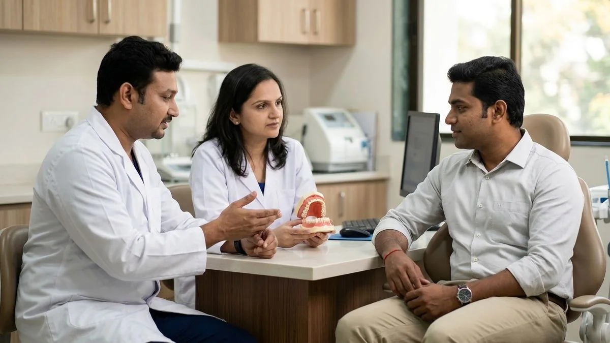
Need expert consultation? Book an appointment with Dr. Pradeep S. or Dr. Kalpa Pandya.
Book AppointmentAlmost everyone has experienced the sharp, irritating pain of a common mouth ulcer. Whether caused by an accidental cheek bite, a stray toothbrush bristle, or a stressful week, these small sores are a routine nuisance. Usually, they fade into memory within a week or two.
However, when a mouth sore persists, grows, or changes texture, it can trigger a deep-seated anxiety: Could this be oral cancer?
Distinguishing between a benign mouth ulcer and the early warning signs of oral cancer is one of the most frequent reasons patients seek our expertise. In our Chennai practice, we frequently consult with patients who have ignored a persistent sore for months, mistakenly believing it to be a simple "heat boil" or "aphthous ulcer."
Understanding the critical differences under the guidance of specialized oral and maxillofacial surgeons can save lives. Early detection remains the single most powerful factor in successfully treating oral malignancies.
Understanding the Common Mouth Ulcer (Aphthous Ulcer)
A standard mouth ulcer, medically termed an aphthous ulcer or canker sore, is a mucosal breach that exposes the underlying nerve endings. This exposure is why even a tiny ulcer can cause significant pain, especially when eating spicy, salty, or acidic foods.
Typical Characteristics of Benign Ulcers
- Appearance: They usually present as round or oval sores with a clean, well-defined yellow or greyish-white center, surrounded by a distinct, bright red inflammatory halo.
- Pain Profile: They are highly painful from the outset, but this pain gradually subsides over several days as the mucosal lining regenerates.
- Location: They develop on mobile, non-keratinized mucosal surfaces, such as the inside of the lips, cheeks, the floor of the mouth, or the soft palate.
- Healing Timeline: They resolve spontaneously within 10 to 14 days without leaving any permanent scars.
Common Triggers
Most benign mouth ulcers are caused by localized, transient factors:
- Minor Trauma: Accidental biting of the cheek or lip, sharp dental restorations, or hard foods.
- Nutritional Deficiencies: Lack of Vitamin B12, iron, folic acid, or zinc.
- Systemic Stress: Emotional or physical stress can alter the local immune response in the oral cavity.
- Hormonal Fluctuations: Frequently observed in women during specific phases of the menstrual cycle.
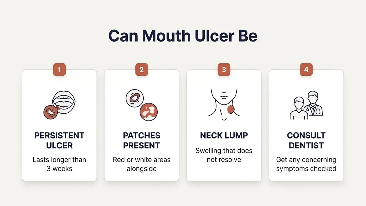
When Does an Ulcer Become Concerning?
While the vast majority of oral sores are benign, certain clinical features demand immediate professional evaluation. In our practice at Mouth Cancer Surgeons, Chennai, we advocate for the Two-Week Rule.
The Two-Week Rule
If any sore, ulcer, red patch, or white lesion in your mouth does not show signs of healing within 14 days, it must be evaluated by a specialist.
Unlike normal tissues, which undergo rapid cellular turnover and heal quickly, cancerous lesions are characterized by uncontrolled, abnormal cellular growth. This dysregulation prevents the body's natural healing mechanisms from repairing the mucosal breach, leaving a chronic, non-healing ulcer.
Clinical Red Flags to Watch For
If your mouth ulcer is accompanied by any of the following symptoms, seek an immediate specialist consultation:
- Painless Progression: Early-stage oral cancer ulcers are frequently painless. The absence of pain often tricks patients into delaying consultation.
- Induration (Hardness): A benign ulcer feels soft and pliable. A cancerous ulcer feels firm, hard, or anchored to the underlying tissues when gently felt (palpated).
- Raised, Rolled, or Everted Edges: While benign ulcers have flat, clean margins, malignant ulcers often exhibit raised, thickened, or rolled-out borders.
- Spontaneous Bleeding: The blood vessels feeding a malignant tumor are fragile and poorly formed. If the ulcer bleeds easily upon light touch, it is a significant warning sign.
- Fixation: The ulcer feels "stuck" to the underlying jawbone or muscle, limiting the normal movement of the tongue or cheek.
Mouth Ulcer vs Oral Cancer Warning Signs to Watch
To help you visualize the clinical differences, we have compiled a comparative overview based on our daily surgical practice.
| Feature | Benign Mouth Ulcer | Oral Cancerous Ulcer |
|---|---|---|
| Pain Level | Sharp, intense pain that decreases over time. | Often painless in early stages; dull, deep ache later. |
| Duration | Heals completely within 10 to 14 days. | Persists beyond 14 days; continues to enlarge. |
| Texture & Feel | Soft, flexible, and tender to the touch. | Hard, firm (indurated), and fixed to tissues. |
| Borders / Edges | Flat, smooth, regular, with a red halo. | Raised, rolled, everted, irregular, or ragged. |
| Bleeding | Rarely bleeds unless actively traumatized. | Bleeds spontaneously or with very light touch. |
| Associated Patches | None, or surrounded by normal pink mucosa. | Often surrounded by persistent red or white patches. |
| Lymph Nodes | Swollen neck nodes are rare (unless infected). | May present with painless, firm, fixed neck swelling. |
Key Warning Signs of Oral Cancer Beyond Ulcers
Oral cancer does not always start as a classic ulcer. It can manifest in several other subtle ways across the oral cavity, including the tongue, buccal mucosa (inner cheek), floor of the mouth, gums (alveolus), and palate.
1. Persistent Red or White Patches
These are known as Oral Potentially Malignant Disorders (OPMD).
- Leukoplakia: Persistent white patches that cannot be scraped off. While many are benign, some undergo malignant transformation. Learn more about managing these lesions on our Oral Precancer (OPMD) page.
- Erythroplakia: Smooth, velvety red patches. These have a significantly higher risk of containing precancerous or early cancerous cells compared to leukoplakia.
- Erythroleukoplakia: A mixed red-and-white patch, which carries the highest risk of malignant transformation.
2. Unexplained Loose Teeth or Sore Gums
If your teeth suddenly become loose without any underlying periodontal (gum) disease, or if a socket fails to heal after a tooth extraction, it could indicate an underlying bone-invasive tumor of the jaw.
3. Difficulty Swallowing, Speaking, or Moving the Jaw
A tumor growing at the base of the tongue, tonsillar region, or deep within the chewing muscles can cause:
- Dysphagia: Pain or difficulty when swallowing.
- Trismus: Inability to open the mouth fully (often associated with Oral Submucous Fibrosis or advanced tumors invading the pterygoid muscles).
- Dysarthria: Slurred speech or difficulty moving the tongue normally.
4. A Lump or Thickening in the Mouth or Neck
A feeling of fullness, a palpable lump in the cheek, or a painless, growing swelling in the neck (cervical lymph node metastasis) is a critical warning sign that requires urgent evaluation.
How We Diagnose Non-Healing Mouth Lesions
When you visit our clinic at Apollo Main Hospital, Greams Road, Chennai, we follow a systematic, evidence-based diagnostic protocol. We understand that this process can be stressful, and we prioritize both clinical accuracy and compassionate patient care.
Step 1: Comprehensive Visual and Physical Examination
Dr. Pradeep S. and Dr. Kalpa Pandya perform a thorough examination of the entire oral cavity, pharynx, and neck. We use high-intensity lighting and specialized palpation techniques to assess the size, depth, texture, and mobility of the lesion, as well as the status of the regional lymph nodes.
Step 2: Diagnostic Biopsy (The Gold Standard)
If a lesion has been present for more than two weeks and exhibits suspicious features, a biopsy is mandatory. A biopsy is the only definitive way to diagnose oral cancer.
- Procedure: We perform an incisional or punch biopsy. Under a small injection of local anesthesia, we remove a tiny, representative sample of tissue from the edge of the ulcer, containing both abnormal and adjacent normal tissue.
- Pathological Review: The tissue is sent to an experienced oral oncopathologist who examines the cells under a microscope to check for dysplasia (precancerous changes) or invasive squamous cell carcinoma.
Step 3: Advanced Imaging (If Malignancy is Confirmed)
If the biopsy confirms oral cancer, we order advanced imaging to determine the stage of the disease:
- Contrast-Enhanced CT (CECT) or MRI: To assess the depth of invasion into surrounding muscles, tissues, and bone.
- PET-CT Scan: To rule out any spread to distant organs and plan precise Oral Cancer Surgery.
The Pathophysiology of Oral Malignancy
To understand why a cancerous ulcer behaves so differently from a standard sore, it helps to understand the underlying biology.
Oral squamous cell carcinoma begins in the thin, flat squamous cells that line the inside of the mouth. Chronic exposure to carcinogens—such as tobacco, alcohol, or mechanical irritation—damages the DNA of these cells. Over time, these genetic mutations cause the cells to multiply uncontrollably.
As the tumor grows, it outstrips its local blood supply, causing the tissue in the center of the lesion to die (necrose). This necrosis creates the ulcerated crater. At the same time, the cancer cells invade deeper tissue layers, recruiting new, fragile blood vessels (neovascularization) and inducing a dense, fibrous reaction around the tumor. This deep invasion is what makes the ulcer feel hard (indurated) and fixed, unlike a superficial, soft aphthous ulcer.
Why Early Detection is Transfomative
The prognosis of oral cancer is directly linked to the stage of the disease at the time of diagnosis. When caught early (Stage I or II), oral cancer is highly treatable, and the five-year survival rate exceeds 80–90% according to global oncological data.
Stage of Diagnosis vs. 5-Year Survival Rate (Approximate)
------------------------------------------------------------
Stage I (Localized, <2cm)       |  85% - 90%  ====================
Stage II (Localized, 2-4cm)     |  70% - 80%  ================
Stage III (Regional lymph nodes)|  40% - 50%  =========
Stage IV (Advanced / Metastatic)|  20% - 30%  ====
------------------------------------------------------------
Early-stage treatment is also much less invasive. It typically involves a localized surgical excision with minimal impact on speech, swallowing, and facial appearance.
Conversely, advanced-stage oral cancers require extensive surgeries, complex jaw reconstructions, and post-operative radiation or chemotherapy. By recognizing the warning signs of a non-healing ulcer early, you can significantly alter the course of treatment and improve your long-term quality of life.
If you are experiencing a persistent mouth sore or have noticed changes in your oral cavity, early consultation is important. Book an appointment with Dr. Pradeep S. and Dr. Kalpa Pandya at Apollo Main Hospital, Greams Road, Chennai.
The Dual-Surgeon Advantage at Mouth Cancer Surgeons
When facing a potential oral cancer diagnosis, the expertise and structure of your surgical team play a critical role in your treatment journey. Our practice, Mouth Cancer Surgeons, is built on a unique, collaborative dual-surgeon model.
Continuous, Coordinated Care
Dr. Pradeep S. and Dr. Kalpa Pandya work together to review, plan, and treat every single patient. From your initial diagnostic biopsy through complex surgical resection, microvascular reconstruction, and long-term surveillance, you are cared for by the same two surgeons. This model ensures complete continuity of care, eliminating communication gaps and providing a highly personalized treatment experience.
- Dr. Pradeep S. brings advanced super-specialty expertise in head & neck surgical oncology and international board-certified maxillofacial reconstructive surgery. He focuses on achieving complete tumor clearance while preserving vital oral functions.
- Dr. Kalpa Pandya brings over a decade of experience in managing oral potentially malignant disorders (OPMD), facial trauma, and complex dental implant rehabilitation. She ensures that your post-cancer rehabilitation allows you to speak, chew, and smile with confidence.
By combining oncological clearance with advanced reconstructive planning from day one, we help our patients achieve the best possible functional and aesthetic outcomes.
Taking Proactive Control of Your Oral Health
While knowing the warning signs is vital, prevention and risk reduction are your best defense against oral cancer.
1. Eliminate Tobacco and Limit Alcohol
Tobacco use remains the leading cause of oral cancer in India. Whether smoked (cigarettes, bidis) or chewed (gutkha, mawa, khaini, betel quid), tobacco introduces potent carcinogens directly to the oral mucosa. When combined with alcohol, the risk multiplies synergistically, as alcohol acts as a solvent, allowing carcinogens to penetrate oral tissues more easily.
2. Address Chronic Dental Irritation
A sharp, chipped tooth, a broken restoration, or an ill-fitting denture that constantly rubs against the tongue or cheek can cause chronic inflammation. Over years, this constant mechanical trauma can trigger cellular changes that may lead to malignancy. Visit your dentist regularly to ensure all surfaces in your mouth are smooth.
3. Maintain Excellent Oral Hygiene
Regular brushing, flossing, and professional dental cleanings reduce chronic inflammation in the oral cavity. Routine dental check-ups also serve as an informal screening, as dentists are trained to spot early mucosal changes that you might not notice.
4. Perform Self-Examinations
Once a month, stand in front of a well-lit mirror and perform a simple self-examination:
- Check your lips and the front of your gums.
- Pull back your cheeks to inspect the inside lining.
- Extend your tongue and look at the top, bottom, and sides.
- Feel the floor of your mouth and along your jawline and neck for any unusual lumps or tenderness.
Seeking Peace of Mind
It is easy to let anxiety take over when you discover a sore in your mouth. However, it is important to remember that most mouth ulcers are entirely harmless. The key is not to panic, but to take structured, proactive action.
If you have an ulcer that has lasted longer than two weeks, do not wait for it to become painful or start bleeding. A simple, quick clinical evaluation with an oral and maxillofacial surgeon can provide definitive answers and peace of mind.
For personalised treatment options and expert care, consult Dr. Pradeep S. and Dr. Kalpa Pandya — Mouth Cancer Surgeons, Chennai. Call +91 96633 03747 or book an appointment.
References
- World Health Organization. "Oral Cancer Prevention and Control." WHO Technical Report Series, 2021.
- National Comprehensive Cancer Network (NCCN). "NCCN Clinical Practice Guidelines in Oncology: Head and Neck Cancers." Version 1.2024, 2024. https://www.nccn.org
- Warnakulasuriya, Saman. "Clinical features of oral potentially malignant disorders: A review." Oral Diseases, 2020. https://doi.org/10.1111/odi.13433
- Indian Council of Medical Research (ICMR). "Consensus Document for the Management of Buccal Mucosa Cancer." ICMR Guidelines, 2022.
- El-Naggar, A. K., et al. "WHO Classification of Head and Neck Tumours." IARC Press, Lyon, 2017.
Authored by

Medically reviewed by