Zygomatic Fracture: Symptoms, Surgery & Recovery
Need expert consultation? Book an appointment with Dr. Pradeep S. or Dr. Kalpa Pandya.
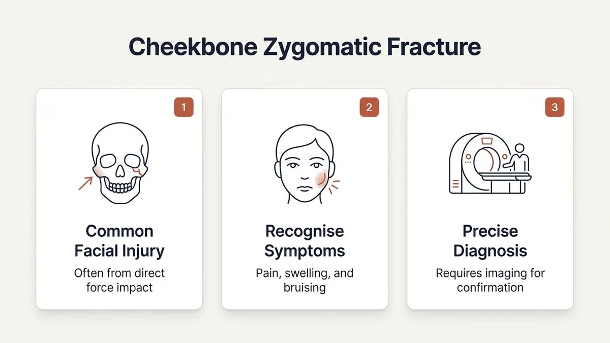
Book AppointmentThe cheekbone, or zygoma, is one of the most prominent features of the human face. It defines our facial contours, supports the eye socket, and plays a crucial role in normal jaw function. Because of its prominent position, it is highly susceptible to high-impact injuries, such as road traffic accidents, sports collisions, physical assaults, or accidental falls.
When this bone breaks, it is medically referred to as a zygomaticomaxillary complex (ZMC) fracture. Understanding cheekbone zygomatic fracture symptoms surgery and recovery is essential for anyone who has sustained a facial injury. Prompt diagnosis and precise surgical intervention by qualified oral and maxillofacial surgeons are vital to restore both facial appearance and critical functions like vision and chewing.
At our practice in Chennai, we regularly manage complex facial trauma. Dr. Pradeep S. and Dr. Kalpa Pandya provide highly specialized, dual-surgeon care at Apollo Main Hospital, Greams Road, ensuring that every patient benefits from their combined expertise from the initial emergency evaluation through to complete reconstructive recovery.
Anatomy of the Cheekbone: Why the Zygoma Matters
The zygoma is not an isolated bone; it is a quadripod structure that connects with four major areas of the skull:
- The Frontal Bone: Forming the outer border of the brow.
- The Maxilla: Forming the upper jaw and the floor of the eye socket (orbit).
- The Temporal Bone: Creating the zygomatic arch that runs toward the ear.
- The Sphenoid Bone: Deep within the skull structure.
Because of these multiple connection points, a severe blow rarely breaks the cheekbone in just one place. Instead, it usually disrupts these structural junctions, leading to what maxillofacial surgeons call a ZMC fracture.
This complex structure sits directly adjacent to several vital anatomical components, including the infraorbital nerve (which provides sensation to your cheek, nose, and upper lip), the extraocular muscles (which control eye movement), and the temporalis muscle (which allows the jaw to close). Consequently, a fracture in this region can have widespread functional implications.
Spotting the Signs: Cheekbone Zygomatic Fracture Symptoms
Following a facial injury, swelling can quickly mask the underlying structural damage. However, several distinct clinical signs point toward a fractured cheekbone. Recognizing these cheekbone zygomatic fracture symptoms surgery and recovery indicators early is crucial for seeking timely specialist care.
1. Facial Flattening and Asymmetry
When the zygoma is fractured and displaced, it typically collapses inward and downward. Once the initial inflammatory swelling subsides, you may notice a distinct loss of projection in the cheek, making one side of the face look flat or sunken compared to the other.
2. Infraorbital Nerve Numbness (Paresthesia)
The infraorbital nerve runs through a small canal in the floor of the eye socket and exits onto the cheek just below the eye. A zygomatic fracture often bruises, stretches, or compresses this nerve. This leads to a distinct "wooden" numbness or tingling sensation in:
- The skin of the affected cheek
- The side of the nose
- The upper lip
- The upper teeth and gums on the injured side
3. Visual Disturbances (Diplopia and Enophthalmos)
Because the cheekbone forms the lateral wall and part of the floor of the eye socket, a fracture can alter the position of the eyeball.
- Double Vision (Diplopia): This occurs if the muscles that move the eye become trapped in the fracture lines or if the orbital volume changes.
- Sunken Eye (Enophthalmos): If the orbit becomes enlarged due to a displaced fracture, the eyeball may sink backward and downward.
4. Restricted Jaw Movement (Trismus)
The zygomatic arch arches over the coronoid process of the lower jaw. If the arch is fractured and pushed inward, it can physically impinge on the jaw bone, preventing you from opening your mouth fully or chewing comfortably.
5. Subconjunctival Hemorrhage and Periorbital Bruising
A classic "black eye" (ecchymosis) accompanied by redness in the white part of the eye (subconjunctival hemorrhage) is highly common. This bleeding occurs because the delicate blood vessels in the eye socket rupture during the impact.
| Symptom | Anatomical Cause | Clinical Significance |
|---|---|---|
| Cheek Flattening | Displacement of the zygomatic body | Aesthetic deformity requiring surgical realignment |
| Numbness in Cheek/Lip | Injury to the infraorbital nerve | Sign of nerve compression; often resolves post-surgery |
| Double Vision (Diplopia) | Entrapment of eye muscles or orbital floor damage | Urgent surgical indicator to prevent permanent vision issues |
| Limited Jaw Opening | Zygomatic arch impinging on the lower jaw | Functional impairment requiring surgical reduction |
| Subconjunctival Bleeding | Rupture of orbital blood vessels | Confirms involvement of the orbital frame |
If you are experiencing any of these symptoms after a facial injury, early consultation is important. Book an appointment with Dr. Pradeep S. and Dr. Kalpa Pandya at Apollo Main Hospital, Greams Road, Chennai.
How Maxillofacial Surgeons Diagnose ZMC Fractures
An accurate diagnosis is the foundation of successful reconstruction. When a patient presents with suspected facial trauma, our surgical team follows a rigorous, multi-step diagnostic protocol.
Clinical Evaluation
During the physical examination, the surgeon gently palpates the facial bones to check for "step-offs"—abrupt unevenness along the bony rims of the eye socket and cheekbone. We also assess eye movements, check for pupillary reflexes, measure jaw opening, and map out areas of sensory loss on the skin.
Advanced 3D Imaging
Standard X-rays are rarely sufficient for complex facial structures. We rely on High-Resolution Computed Tomography (HRCT) with 3D reconstruction. This technology allows us to:
- View the fracture lines from every angle
- Measure the exact degree of bone displacement
- Assess the integrity of the delicate orbital floor
- Plan the precise placement of surgical plates and screws
Surgical Options: How Cheekbone Fractures Are Repaired
Not every cheekbone fracture requires surgery. If the bone is not displaced, your vision is unaffected, your jaw opens normally, and there is no aesthetic deformity, we may recommend conservative management. This involves a soft diet, avoiding physical exertion, and close monitoring with follow-up scans.
However, when displacement occurs, surgical intervention is necessary to prevent long-term functional and cosmetic issues. The gold standard for repair is Open Reduction and Internal Fixation (ORIF).
The ORIF Procedure
Under general anesthesia, the surgeon accesses the fractured bone through carefully planned incisions designed to minimize visible scarring.
- Incisions: These are often placed inside the upper mouth (intraoral), within the natural crease of the eyelid (subciliary or transconjunctival), or within the hairline.
- Reduction: Using specialized instruments, the displaced cheekbone is gently guided back into its precise anatomical position.
- Fixation: Once aligned, the bones are secured using biocompatible, ultra-thin titanium plates and micro-screws. These implants are highly stable, well-tolerated by the body, and typically remain in place permanently without causing any irritation.
For comprehensive details on how we approach these complex procedures, you can read more about our specialized approach to facial trauma and emergency surgery and learn about the techniques used in open reduction and internal fixation (ORIF).
What to Expect During Cheekbone Zygomatic Fracture Recovery
The recovery phase is just as critical as the surgery itself. Understanding what to expect day-by-day helps reduce anxiety and ensures a smooth healing process.
Immediately Post-Surgery (Days 1 to 3)
- Swelling and Bruising: Peak swelling typically occurs 48 to 72 hours after surgery. Applying cold compresses to the area for 15 minutes at a time can help minimize discomfort.
- Pain Management: Mild to moderate pain is expected and is managed effectively with prescribed analgesics and anti-inflammatory medications.
- Hospital Stay: Most patients undergoing uncomplicated ORIF for a cheekbone fracture are discharged within 24 to 48 hours.
The First Two Weeks
- Nose Blowing Warning: You must not blow your nose for at least 4 to 6 weeks. The cheekbone forms the wall of the maxillary sinus. Blowing your nose can force air and bacteria from the sinus cavity into the surrounding facial tissues and eye socket, causing severe swelling or infection.
- Dietary Restrictions: To avoid putting pressure on the newly repaired bones, you must adhere to a strict soft-food diet (e.g., porridges, mashed potatoes, soups, and yogurts) for the first 2 weeks, gradually transitioning to soft solid foods as guided by your surgeon.
- Oral Hygiene: Keeping the mouth clean is vital, especially if intraoral incisions were used. Gentle rinsing with warm salt water or a prescribed chlorhexidine mouthwash after meals is highly recommended.
Long-Term Healing (Weeks 3 to 8)
- Bone Consolidation: While you will begin to feel much better by week 3, the bones take approximately 6 to 8 weeks to regain their full structural strength.
- Activity Restrictions: Light daily activities can be resumed within a week, but strenuous exercise, heavy lifting, and any activities that carry a risk of facial impact (such as contact sports) must be avoided for at least 2 months.
Potential Complications of Untreated Cheekbone Fractures
Choosing to delay or bypass treatment for a displaced cheekbone fracture can lead to permanent functional deficits and aesthetic deformities that become significantly harder to correct later.
[Displaced Cheekbone Fracture]
       │
       ├─► No Treatment ──► Permanent Facial Asymmetry & Sunken Eye (Enophthalmos)
       │
       ├─► Delayed Care ──► Chronic Double Vision (Diplopia) & Nerve Damage
       │
       └─► Expert ORIF  ──► Restored Facial Structure, Normal Vision & Jaw Function
Chronic Facial Asymmetry
Once a fractured cheekbone heals in a displaced position (malunion), the flat appearance of the cheek becomes permanent. Correcting a malunion later requires complex secondary osteotomies (intentionally re-breaking the bone) or custom-made facial implants.
Persistent Diplopia and Vision Problems
If the orbital floor remains damaged or the eye muscles remain entrapped, double vision can become permanent, severely impacting your ability to drive, read, or perform daily tasks safely.
Permanent Facial Numbness
Prolonged compression of the infraorbital nerve can lead to irreversible nerve damage, resulting in permanent numbness or chronic neuropathic pain across the cheek and upper lip.
Why Choose Mouth Cancer Surgeons for Facial Trauma in Chennai
While our practice brand is "Mouth Cancer Surgeons," our core specialization is rooted deeply in Oral & Maxillofacial Surgery. Reconstructing complex facial structures after trauma requires the exact same high-level surgical precision, structural understanding, and aesthetic consideration as reconstructing the face after major tumor resections.
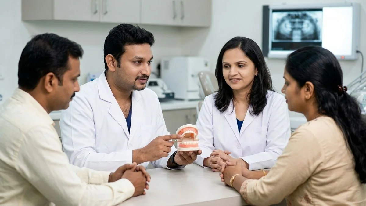
- Dual-Surgeon Expertise: Every complex trauma case is evaluated and planned by both Dr. Pradeep S. and Dr. Kalpa Pandya. Having two highly trained maxillofacial specialists review your scans and coordinate your care ensures optimal structural and aesthetic outcomes.
- Comprehensive Care Model: From the moment you are admitted to Apollo Main Hospital, Greams Road, through your surgery and long-term follow-up visits, you are cared for by the exact same two surgeons. We do not delegate your critical recovery phases to junior staff.
- Advanced Reconstruction Techniques: We utilize state-of-the-art 3D imaging, virtual surgical planning, and high-quality titanium plating systems to reconstruct the facial skeleton with extreme precision, prioritizing hidden incisions to minimize visible scarring.
If you or a loved one has sustained a facial injury and are seeking expert guidance on cheekbone zygomatic fracture symptoms, surgery, and recovery, our team is here to provide world-class, compassionate care.
For personalised treatment options and expert care, consult Dr. Pradeep S. and Dr. Kalpa Pandya — Mouth Cancer Surgeons, Chennai. Call +91 96633 03747 or book an appointment.
References
- Ellis, Edward, and Bruce E. Kullman. "Association of partner fractures with zygomaticomaxillary complex fractures." Journal of Oral and Maxillofacial Surgery, 2013.
- Strong, E. Bradley, and Robert M. Kellman. "Zygomaticomaxillary Complex Fractures." Facial Plastic Surgery Clinics of North America, 2021.
- Neuman, Brian J., et al. "Evaluation and Management of Zygomatic Arch Fractures." The Journal of Craniofacial Surgery, 2018.
- Indian Association of Oral and Maxillofacial Surgeons (AOMSI). "Clinical Practice Guidelines for Management of Maxillofacial Trauma." Journal of Maxillofacial and Oral Surgery, 2020.
- National Institutes of Health (NIH). "Surgical Management of Midfacial and Zygomatic Fractures: A Review of Current Protocols." PMC Craniofacial Medicine, 2022.
Authored by

Medically reviewed by