Oral Cancer Screening & Self-Exam Guide | Chennai Expert
Need expert consultation? Book an appointment with Dr. Pradeep S. or Dr. Kalpa Pandya.
Book AppointmentOral cancer is one of the most prevalent malignancies in India, particularly across Tamil Nadu and South India, largely driven by the widespread use of tobacco, betel nut (paan), and alcohol. Despite its high incidence, a vast majority of cases are diagnosed at advanced stages (Stage III or IV), where treatment becomes highly complex and survival rates decline.
Implementing routine oral cancer screening and self-examination early detection protocols can fundamentally change this trajectory. When oral squamous cell carcinoma or its precursor lesions are identified early, treatment is far less invasive, facial aesthetics and oral functions are preserved, and long-term cure rates exceed 80–90%.
At Mouth Cancer Surgeons in Chennai, our surgical team—consisting of Dr. Pradeep S. and Dr. Kalpa Pandya—frequently manages patients who could have avoided extensive jaw reconstructions if their lesions had been detected just a few months earlier. This comprehensive guide outlines the clinical importance of screening, provides a detailed step-by-step protocol for performing a self-examination at home, and explains when you must seek an urgent specialist evaluation.
Why Early Detection of Oral Cancer is a Lifesaver
The clinical progression of oral cancer is highly predictable, often starting as a localized, superficial change in the oral mucosa before invading deeper muscle, bone, and regional lymph nodes in the neck. Catching the disease while it is still localized is the single most important factor in determining a patient's prognosis.
Survival Rates and Clinical Outcomes
Data from the National Cancer Institute and global oncological registries demonstrate a stark contrast in survival rates based on the stage at diagnosis:
- Localized Disease (Stage I & II): The 5-year relative survival rate is approximately 85% to 90%. At this stage, the tumor is small (less than 4 cm) and has not spread to the lymph nodes.
- Regional Spread (Stage III & IV with neck node involvement): The 5-year survival rate drops to 40% to 50%.
- Distant Metastasis: If the cancer spreads to distant organs, the 5-year survival rate falls below 20%.
Minimizing the Need for Major Reconstructive Surgery
When oral cancer is diagnosed late, surgical management typically requires a composite resection. This often involves removing a segment of the jawbone (mandible or maxilla), a large portion of the tongue, or the inner cheek lining, followed by a complex microvascular free flap reconstruction.
Conversely, early-stage lesions can frequently be treated with wide local excisions or minimally invasive transoral surgeries. This preserves natural swallowing, speech, and facial appearance, avoiding the physical and psychological toll of extensive reconstructive procedures.
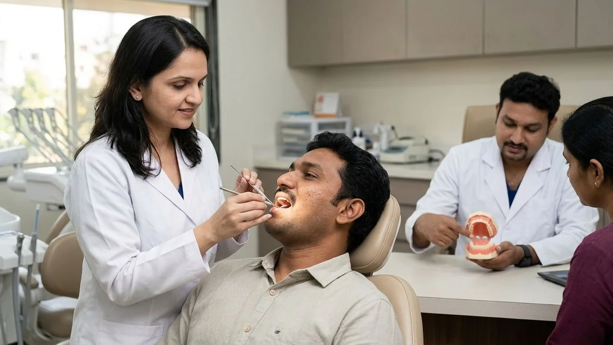
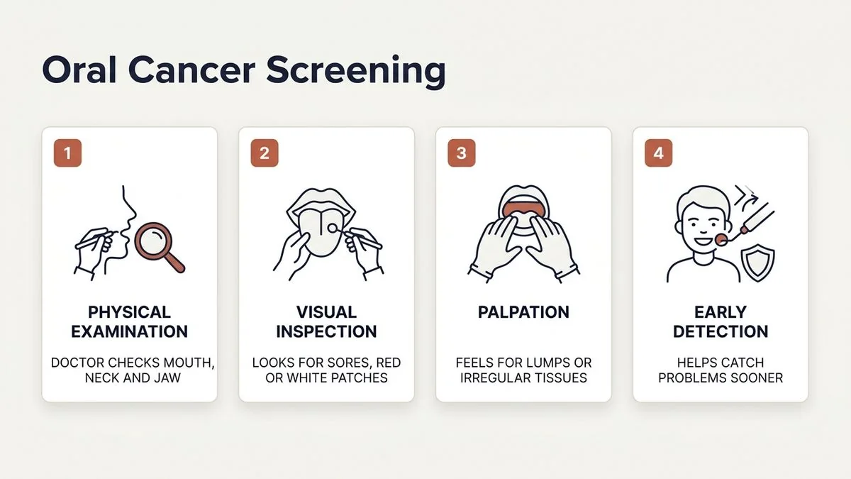
What is Professional Oral Cancer Screening?
A professional oral cancer screening is a rapid, painless, and highly effective clinical examination performed by a specialist, such as an oral & maxillofacial surgeon. In our practice at Apollo Main Hospital, Greams Road, Chennai, a comprehensive screening involves two primary components:
1. Visual Inspection
Using high-intensity, color-corrected illumination and dental mirrors, the surgeon systematically examines every square millimeter of your oral cavity. This includes the lips, the inside of the cheeks (buccal mucosa), the gums (alveolar ridges), the roof of the mouth (hard and soft palate), the back of the throat (oropharynx), and all surfaces of the tongue.
2. Bimanual Palpation
Many early cancers or precancerous changes present as deep-seated induration (firmness) beneath an otherwise normal-looking mucosal surface. The surgeon uses gloved hands to feel the floor of the mouth, the tongue, and the cheeks, pressing from both the inside and outside simultaneously. We also perform a comprehensive neck examination to check for enlarged, firm, or fixed lymph nodes.
Step-by-Step Guide to Oral Cancer Self-Examination at Home
Performing a self-examination at home takes less than five minutes and should be done once a month. This practice helps you become familiar with the normal anatomy of your mouth, making it easy to spot any new or unusual changes immediately.
Setting Up: What You Need
- A brightly lit room with a large wall mirror.
- A small, bright flashlight or a smartphone light.
- A clean piece of gauze or a small tissue to help hold your tongue.
- Thoroughly washed hands.
The 7-Step Self-Exam Protocol
Follow this structured sequence to ensure you do not miss any area of your mouth:
Step 1: Examine the Lips
Look at your lips in the mirror with your mouth closed, checking for changes in color, texture, or symmetry. Next, open your mouth, gently pull your upper lip upward, and pull your lower lip downward to inspect the inner pink lining for any red or white patches, ulcers, or rough spots.
Step 2: Check the Inner Cheeks (Buccal Mucosa)
Using your thumb and index finger, gently pull one cheek outward. Use your flashlight to inspect the entire inner lining. Look for red, white, or dark spots, ulcers, or any areas that look raw. Run your index finger along the inside of the cheek to feel for lumps, bumps, or areas of unusual thickness or tenderness. Repeat this process on the other cheek.
Step 3: Inspect the Gums (Alveolar Ridges)
Smile widely to expose your gums. Inspect the outer gums surrounding both your upper and lower teeth, looking for swelling, persistent redness, or ulcerations. Run your finger along your gums to feel for any rough texture or firm masses.
Step 4: Examine the Tongue (All Surfaces)
Extend your tongue as far as possible and inspect the top surface (dorsum) for changes in color, loss of the normal velvety texture, or unusual coatings.
Next, wrap a clean piece of gauze or tissue around the tip of your tongue to hold it gently. Pull the tongue to the far right side and inspect the lateral border. This lateral border of the tongue is one of the most common sites for oral cancer. Repeat this process by pulling the tongue to the far left side. Finally, touch the tip of your tongue to the roof of your mouth to inspect the underside (ventral surface).
Step 5: Check the Floor of the Mouth
With your tongue still raised, shine your light onto the floor of your mouth. Look closely for any ulcers, red or white patches, or swelling. Place one index finger inside your mouth under your tongue, and place the fingers of your other hand on the skin under your jaw. Gently press the two hands together to feel for any deep lumps, nodules, or firm areas.
Step 6: Inspect the Roof of the Mouth (Palate)
Tilt your head back and open your mouth wide. Shine your light onto the roof of your mouth to inspect both the hard palate (front part) and the soft palate (back part). Look for changes in color, ulcers, or asymmetric swellings. Run your index finger over the area to check for any lumps or rough patches.
Step 7: Palpate the Neck and Jawline
Using the pads of your fingers, feel along both sides of your neck, under your jawline, and around your collarbone. Press gently but firmly, feeling for any enlarged, hard, or tender lymph nodes (lumps) that feel different on one side compared to the other.
Distinguishing Normal Tissue from Warning Signs
It is common to find anatomical variations in the mouth that are completely harmless. Understanding the difference between normal oral structures and suspicious lesions can help prevent unnecessary anxiety while ensuring you seek help when it is truly needed.
| Oral Feature / Structure | Normal / Benign Presentation | Suspicious / Malignant Signs |
|---|---|---|
| Color of Mucosa | Uniformly pink, moist, and smooth. | Persistent red patches (erythroplakia), bright white patches (leukoplakia), or mixed red-and-white areas. |
| Mouth Ulcers | Small, painful sores (like aphthous ulcers) that heal completely within 10 to 14 days. | Ulcers that do not heal after two weeks, are painless, have raised, firm borders, or bleed easily when touched. |
| Tissue Texture | Soft, pliable, and symmetrical on both sides. | Areas that feel hard, thick, or fixed to the underlying tissue (induration). |
| Tongue Anatomy | Symmetrical papillae on the top surface; prominent, blue-purple veins on the underside. | Asymmetric lumps, deep cracks, or raw, red areas on the sides or underside of the tongue. |
| Bony Prominences | Smooth, hard, slow-growing lumps in the middle of the palate or inside the lower jaw (tori). | Rapidly growing bony swellings, loose teeth with no obvious dental cause, or unexplained jaw pain. |
Identifying Oral Potentially Malignant Disorders (OPMDs)
Many oral cancers do not appear suddenly; they often develop from pre-existing conditions known as Oral Potentially Malignant Disorders (OPMDs). If you notice any of the following conditions during your self-examination, you should have them evaluated at our oral precancer (OPMD) clinic:
- Leukoplakia: A white patch or plaque that cannot be rubbed off or characterized clinically as any other disease.
- Erythroplakia: A fiery red, velvety patch that has a high risk of transforming into cancer.
- Oral Submucous Fibrosis (OSMF): A chronic condition common in betel nut chewers, characterized by progressive stiffness of the oral mucosa, a burning sensation when eating spicy food, and a gradual loss of the ability to open the mouth fully.
High-Risk Groups Who Require Regular Screenings
While oral cancer can affect anyone, certain lifestyle factors and viral exposures significantly increase your risk. If you fall into any of the following high-risk categories, routine professional screenings and monthly self-examinations are highly recommended:
- Tobacco Users: This includes smoking cigarettes, bidis, or cigars, as well as using smokeless tobacco products like gutkha, khaini, and mawa. Tobacco contains potent carcinogens that damage the DNA of your oral mucosal cells.
- Betel Nut (Areca Nut) Chewers: Betel nut is a known Group 1 human carcinogen. It causes chronic irritation and mucosal damage, often leading to OSMF and oral cancer.
- Heavy Alcohol Consumers: Alcohol acts as a solvent, making the delicate oral mucosa more permeable to other carcinogens, such as those found in tobacco.
- HPV Exposure: Infection with high-risk strains of the Human Papillomavirus (specifically HPV-16 and HPV-18) is a major risk factor for cancers of the tonsils, base of the tongue, and throat (oropharyngeal cancer).
- Previous Cancer Survivors: Individuals who have previously been treated for oral, head, or neck cancer have a significantly higher risk of developing a second primary tumor in the oral cavity.
When to Transition from Self-Examination to Professional Consultation
If you find something unusual during your self-examination, try not to panic. Many oral lesions are benign or related to minor dental issues, such as a sharp tooth rubbing against your cheek. However, you should schedule an urgent professional evaluation if you experience any of the following warning signs:
- The Two-Week Rule: Any mouth ulcer, red or white patch, or rough spot that does not heal or improve within 14 days, even after removing potential irritants like sharp teeth or ill-fitting dentures. For more details on identifying these warning signs, read our article on how to tell if a mouth ulcer is cancerous.
- Unexplained Bleeding: Persistent bleeding in the mouth without an obvious cause, such as active gum disease.
- Numbness or Loss of Sensation: A sudden, unexplained loss of feeling in your lip, tongue, or gums.
- Difficulty with Basic Functions: Pain or stiffness when chewing, swallowing, speaking, or moving your jaw or tongue.
- A Persistent Neck Lump: Any firm, painless lump in your neck that remains present for more than two weeks.
If you are experiencing any of these symptoms, early consultation is important. Book an appointment with Dr. Pradeep S. and Dr. Kalpa Pandya at Apollo Main Hospital, Greams Road, Chennai.
What Happens If an Abnormality is Found?
If our surgical team identifies a suspicious lesion during your clinical screening, we will guide you through a clear, step-by-step diagnostic process.
1. Diagnostic Biopsy
A biopsy is the only definitive way to diagnose oral cancer. It is a quick, safe, and minor procedure performed under local anesthesia in our clinic. A tiny sample of tissue is taken from the suspicious area and sent to an experienced oral pathologist for analysis.
2. Advanced Imaging and Staging
If the biopsy confirms the presence of precancerous cells or cancer, we will order imaging studies, such as a contrast-enhanced CT scan or an MRI. These scans help us determine the exact size of the lesion and check if it has affected nearby bone or lymph nodes in the neck.
3. Personalized Treatment Planning
At Mouth Cancer Surgeons, every patient benefit from our dual-surgeon model. Dr. Pradeep S. and Dr. Kalpa Pandya personally review every case together. We work closely with a multidisciplinary oncology board at Apollo Main Hospital to design a personalized treatment plan.
Depending on the stage of the disease, treatment may involve a wide surgical excision, reconstructive surgery, or a combination of surgery, radiation, and chemotherapy. To learn more about our comprehensive approach, visit our multidisciplinary oncology care page.
Why Choose Mouth Cancer Surgeons in Chennai?
Choosing the right surgical team is critical when dealing with oral cancer or precancerous conditions. Here is what sets our practice apart:
- Dual-Surgeon Expertise: Our patients receive coordinated care from two highly trained, dedicated oral & maxillofacial surgeons. Dr. Pradeep S. and Dr. Kalpa Pandya collaborate on every case, from the initial consultation and biopsy through surgery, reconstruction, and long-term follow-up.
- Comprehensive Care: We specialize in the entire spectrum of oral and maxillofacial care. This includes screening, managing OPMDs, performing complex cancer resections, and carrying out advanced reconstructive surgeries.
- State-of-the-Art Facilities: Our primary surgical practice is based at Apollo Main Hospital, Greams Road, Chennai. This allows us to offer our patients access to world-class operating theaters, advanced diagnostic imaging, and dedicated intensive care units.
Monthly self-examinations and regular professional screenings are simple, life-saving habits. Taking just five minutes once a month to check your mouth can help you catch potential issues early, protecting your health, your smile, and your future.
For personalised treatment options and expert care, consult Dr. Pradeep S. and Dr. Kalpa Pandya — Mouth Cancer Surgeons, Chennai. Call +91 96633 03747 or book an appointment.
References
- National Cancer Institute. "Oral Cavity and Pharyngeal Cancer — Cancer Stat Facts." SEER Database, 2023. https://seer.cancer.gov/statfacts/html/oralcav.html
- Warnakulasuriya, Saman. "Clinical features and presentation of oral potentially malignant disorders." Oral Oncology, 2020.
- Indian Council of Medical Research (ICMR). "Consensus Document for Management of Buccal Mucosa Cancer." ICMR Guidelines, 2019.
- National Comprehensive Cancer Network (NCCN). "NCCN Clinical Practice Guidelines in Oncology: Head and Neck Cancers." NCCN, 2024. https://www.nccn.org
- Brocklehurst, Paul, et al. "Screening programmes for the early detection and prevention of oral cancer." Cochrane Database of Systematic Reviews, 2013.
Authored by

Medically reviewed by