Cheek Cancer Symptoms, Surgery & Recovery | Chennai
Need expert consultation? Book an appointment with Dr. Pradeep S. or Dr. Kalpa Pandya.
Book AppointmentA persistent, painless ulcer on the inside of the cheek is frequently misdiagnosed or ignored as a simple aphthous ulcer, an accidental bite, or minor trauma from a sharp tooth. However, when an ulcer or patch on the inner cheek lining fails to heal within two to three weeks, it requires urgent evaluation by a specialist. This lining is anatomically known as the buccal mucosa, and cancers arising here are highly aggressive, requiring prompt, multi-specialty intervention.
In our practice at Mouth Cancer Surgeons in Chennai, we frequently consult with patients who have ignored early warning signs, allowing the disease to progress to advanced stages. Understanding the timeline of buccal mucosa cheek cancer symptoms surgery and recovery is crucial for patients and caregivers facing this diagnosis.
Through this guide, Dr. Pradeep S. and Dr. Kalpa Pandya outline the clinical presentation, advanced surgical interventions, and recovery protocols that define modern, high-quality oral cancer care.
What is Buccal Mucosa Cheek Cancer?
The buccal mucosa refers to the inner mucosal lining of the cheeks and lips, extending from the line of attachment of the mucosa to the alveolar ridges (gums) to the pterygomandibular raphe (the back of the cheek).
Buccal mucosa cancer is a subtype of oral cavity cancer surgery and is predominantly diagnosed as Oral Squamous Cell Carcinoma (OSCC). Squamous cells are the flat, scale-like cells that form the outermost layer of the oral mucosa. When these cells undergo genetic mutations—often driven by chronic exposure to carcinogens—they begin to multiply uncontrollably, forming a localized tumor that can eventually invade deep muscle tissue, adjacent jawbones, and regional lymph nodes in the neck.
In India and across South Asia, buccal mucosa cancer is exceptionally common, accounting for a massive share of all diagnosed oral malignancies. This high incidence is directly linked to cultural habits, specifically the chewing of smokeless tobacco, betel quid (paan), and areca nut, which are held in the buccal sulcus (the pocket between the cheek and teeth) for prolonged periods.
Recognizing Buccal Mucosa Cheek Cancer Symptoms
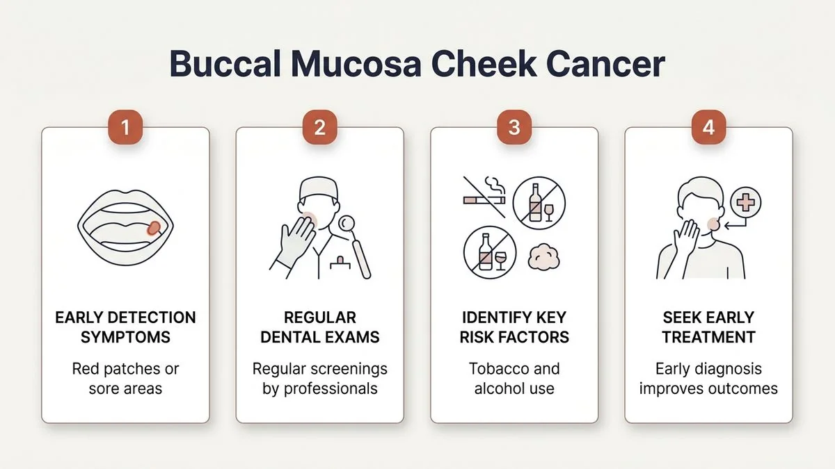
Early detection is the single most important factor determining the success of treatment. Patients must look out for localized changes in the mouth that do not resolve with standard topical ointments or dental care.
Early Warning Signs
- The Non-Healing Ulcer: A sore on the inner cheek that does not heal within 14 days. Unlike common canker sores, a malignant ulcer is often painless in its early stages, has raised, hardened (indurated) borders, and may bleed easily when touched. Learn more about identifying these signs in our guide on warning signs of a malignant mouth ulcer.
- White or Red Patches: Persistent white patches (leukoplakia) or velvety red patches (erythroplakia) on the inner cheek. These are often classified as oral potentially malignant disorders (OPMD) and have a high rate of malignant transformation.
- Localized Thickening: Feeling a distinct lump, thickness, or "fullness" inside the cheek with the tongue or finger, even if no visible ulcer is present.
Advanced Symptoms
- Trismus (Difficulty Opening the Mouth): As the tumor grows, it penetrates deep into the buccinator muscle or the masticatory muscles (such as the medial pterygoid or masseter). This causes progressive scarring and muscle spasm, severely restricting the patient's ability to open their mouth.
- Pain and Referred Otalgia: Pain that originates in the cheek but radiates to the ear on the same side. This occurs when the tumor infiltrates deep sensory nerves, such as the mandibular branch of the trigeminal nerve.
- Mobility of Teeth or Jaw Pain: If the tumor spreads inward, it can invade the upper jaw (maxilla) or lower jaw (mandible), causing teeth to loosen or causing deep, dull bone pain.
- Neck Lumps: A firm, painless swelling in the neck, which indicates that the cancer has metastasized to the cervical lymph nodes.
| Symptom Stage | Clinical Presentation | Patient Experience | Action Required |
|---|---|---|---|
| Early Stage | Superficial ulcer, white/red patch, mild induration | Painless, feels like a rough spot or a minor bite | Immediate clinical biopsy |
| Intermediate Stage | Deep ulceration, infiltration of buccinator muscle | Mild pain, early restriction in mouth opening | Diagnostic staging and surgical planning |
| Advanced Stage | Bone invasion, skin involvement, neck node metastasis | Severe pain, earache, severe trismus, visible neck lump | Multidisciplinary oncology care and major reconstructive surgery |
If you or a loved one is experiencing any of these symptoms, early consultation is important. Book an appointment with Dr. Pradeep S. and Dr. Kalpa Pandya at Apollo Main Hospital, Greams Road, Chennai.
The Causes and Risk Factors
The primary driver of buccal mucosa cancer is chronic mucosal irritation. The delicate squamous epithelium of the inner cheek is not designed to withstand constant chemical and physical trauma.
- Smokeless Tobacco and Areca Nut: Placing betel quid, gutkha, khaini, or paan masala in the cheek pocket exposes the mucosal cells to highly carcinogenic nitrosamines. This leads to chronic inflammation, localized fibrosis, and cellular DNA damage.
- Smoking and Alcohol Consumption: Cigarette, bidi, or cigar smoke introduces heat and chemical carcinogens. When combined with alcohol—which acts as a solvent, making the mucosal membrane more permeable—the risk of malignant transformation increases exponentially.
- Chronic Dental Trauma: A sharp, broken, or decayed tooth, or an ill-fitting dental denture that constantly rubs against the inner cheek, can create a chronic ulcer. Over months or years, the continuous cycle of cellular damage and rapid repair can trigger genetic mutations leading to cancer.
Diagnosis and Staging
When a patient presents with a suspicious lesion on the inner cheek, a systematic diagnostic protocol is initiated to confirm the diagnosis and map the extent of the disease.
1. Clinical Examination and Biopsy
The gold standard for diagnosing buccal mucosa cancer is an incisional biopsy. Under local anesthesia, a small tissue sample is taken from the edge of the ulcer or patch and sent to a neuropathologist/oral pathologist. This confirms whether the lesion is malignant and determines its histological grade (well, moderately, or poorly differentiated).
2. Radiographic Imaging
To plan the surgery, we must know precisely how deep the tumor has penetrated:
- Contrast-Enhanced CT (CECT) or MRI: These scans show the depth of invasion into the cheek muscles, involvement of the masticator space, and whether the tumor has spread to the lymph nodes in the neck.
- Orthopantomogram (OPG) or Cone Beam CT (CBCT): Used to assess if the tumor has eroded the bone of the lower or upper jaw.
- PET-CT Scan: Typically reserved for advanced stages to rule out distant metastasis (spread to the lungs, liver, or bones) and to assess overall disease burden.
Surgical Treatment for Buccal Mucosa Cancer
Surgery is the cornerstone of curative treatment for buccal mucosa cancer. The primary goal is to completely remove the cancer while preserving as much oral function and facial aesthetics as possible.
Because buccal mucosa tumors can grow rapidly and spread to adjacent tissues, the surgical plan must address three key areas: the primary tumor, the jawbone (if involved), and the lymph nodes in the neck.
1. Wide Local Excision
The surgeon removes the entire visible tumor along with a 1.5 to 2-centimeter margin of healthy-looking tissue around it. This "clear margin" is essential to ensure that microscopic cancer cells left at the boundaries do not cause a recurrence. Depending on the tumor's depth, this excision may include the underlying buccinator muscle and, in advanced cases, may extend to involve the overlying skin of the cheek.
2. Management of the Jaw (Mandibulectomy or Maxillectomy)
If the cancer is close to or invading the jawbone, a portion of the bone must be removed:
- Marginal Mandibulectomy: If the tumor is adjacent to the lower jaw but has not invaded the bone marrow, the surgeon shaves off a rim of bone, keeping the continuity of the jaw intact.
- Segmental Mandibulectomy: If the cancer has invaded deep into the lower jawbone, a complete segment of the mandible must be removed. This requires immediate reconstruction to prevent the jaw from collapsing.
3. Neck Dissection
Even if neck scans show no obvious signs of cancer, buccal mucosa tumors have a high rate of microscopic spread to the lymph nodes in the neck. A neck dissection is performed to remove these lymph nodes (usually Levels I, II, and III for early stages, and Levels I-V for advanced stages). This is both a therapeutic and a staging procedure, helping us determine if further treatment like radiation is needed.
4. Reconstructive Surgery
Removing a large portion of the inner cheek, and potentially parts of the jaw and skin, leaves a significant defect. Without reconstruction, the patient would suffer from severe facial disfigurement, an inability to open their mouth, and a loss of the ability to speak or swallow.
Modern oral oncology relies heavily on reconstructive and restorative surgery. At Mouth Cancer Surgeons, we specialize in advanced microvascular free flap reconstruction:
- Anterolateral Thigh (ALT) Flap: Taking skin, fat, and fascia from the thigh along with its blood vessels, and transplanting it to the cheek defect. The blood vessels are micro-surgically sutured to blood vessels in the neck to restore circulation.
- Radial Forearm Free Flap: Ideal for thinner, more pliable reconstructions of the inner cheek lining, allowing for excellent flexibility and movement.
- Fibula Free Flap: If a segment of the jawbone was removed, the fibula bone from the lower leg, along with its skin and blood vessels, is used to rebuild a functional, strong jawbone.
What to Expect During Recovery
Recovery after buccal mucosa surgery is a gradual process that requires professional guidance, patience, and dedicated rehabilitation.
The Immediate Post-Operative Phase (Hospital Stay: 7–14 Days)
- Airway Management: In major surgeries, especially those involving jaw reconstruction or neck dissection, a temporary breathing tube in the windpipe (tracheostomy) may be placed to ensure a safe, secure airway while facial swelling subsides. This is usually removed within 5 to 7 days.
- Nutrition: Patients cannot eat through the mouth immediately after surgery to allow the internal cheek grafts to heal without irritation. Nutrition is delivered via a nasogastric (Ryle's) tube directly into the stomach.
- Flap Monitoring: If a microvascular free flap was used, our surgical team monitors the graft every hour for the first 48 to 72 hours using clinical checks and Doppler ultrasound to ensure optimal blood flow.
The Intermediate Phase (Weeks 2–6)
- Wound Healing and Suture Removal: External skin sutures are typically removed 7 to 10 days after surgery. The internal mucosal lining heals completely within 3 to 4 weeks.
- Transitioning to Oral Intake: Once the surgical team confirms that the internal grafts have healed and there are no leaks, the patient is transitioned from liquid diets to soft, pureed foods, gradually working back to a normal diet.
- Jaw Exercises: To combat trismus (jaw stiffness), patients must perform structured jaw-opening exercises daily. This prevents the surgical scar tissue from contracting and locking the jaw.
Long-Term Rehabilitation (Months 2–6 and Beyond)
- Speech and Swallowing Therapy: Working with specialized speech pathologists helps patients adapt to the reconstructed tissues, ensuring clear speech and safe swallowing.
- Dental Rehabilitation: Once the surgical site is completely stable, patients who lost teeth or parts of the jaw can undergo dental implant placement to restore full chewing function. Learn more about post-cancer dental rehabilitation on our dental implants and pre-prosthetic surgery page.
| Recovery Phase | Timeline | Primary Focus | Key Milestones |
|---|---|---|---|
| Phase 1: Acute | Days 1 to 14 | Airway safety, graft monitoring, wound healing, pain control | Discharge from hospital, removal of tracheostomy |
| Phase 2: Transition | Weeks 2 to 6 | Transitioning to soft oral diet, starting jaw-stretching exercises | Removal of feeding tube, independent oral intake |
| Phase 3: Functional | Months 2 to 6 | Speech therapy, swallowing coordination, scar remodeling | Return to clear speech, near-normal jaw opening |
| Phase 4: Restorative | Month 6 onwards | Long-term surveillance, dental implant rehabilitation | Complete functional restoration, regular cancer screening |
Multidisciplinary Care and Adjuvant Therapy
For many patients, surgery is only the first step. Once the removed tumor and lymph nodes are analyzed by a pathologist, we determine if additional treatment is required to prevent the cancer from returning.
If the pathology report reveals high-risk features—such as cancer cells close to the surgical margins, spread to multiple neck lymph nodes, or invasion into local nerves (perineural invasion)—the patient will be referred for adjuvant therapy:
- Radiation Therapy: High-energy X-rays targeted at the surgical site and neck to destroy any microscopic cancer cells that may remain.
- Chemoradiotherapy: Combining radiation with low-dose chemotherapy to make the cancer cells more sensitive to the radiation.
At Mouth Cancer Surgeons, we coordinate this seamlessly through our multidisciplinary oncology care network, ensuring our patients receive world-class, integrated treatment.
Prognosis and Survival Rates
The prognosis for buccal mucosa cancer is highly dependent on the stage at diagnosis. According to clinical data, patients diagnosed with Stage I or II cheek cancer have an encouraging 5-year survival rate of approximately 75% to 80% when treated with definitive surgery. However, for advanced stages (Stage III and IV), the survival rate declines, highlighting the critical importance of early diagnosis. For a deeper understanding of prognosis and outcomes, read our article on is mouth cancer curable.
The Dual-Surgeon Model: Why Choose Us?
When facing a complex diagnosis like buccal mucosa cancer, the experience and structure of your surgical team make a profound difference in your treatment outcome.
At Mouth Cancer Surgeons in Chennai, we operate under a unique dual-surgeon model.
- Dr. Pradeep S. (MDS, FHNO, FIBCSOMS) is an internationally board-certified head and neck surgical oncologist who specializes in complex cancer resections and advanced microvascular reconstructions.
- Dr. Kalpa Pandya (MDS, FHNS) brings over a decade of specialized experience in managing oral potentially malignant disorders (OPMDs), facial trauma, and comprehensive dental implant rehabilitation.
Why this model benefits you:
- Simultaneous Surgery: During major reconstructive procedures, both surgeons work together in the operating theatre. While one surgeon focuses on safely removing the tumor (resection), the other simultaneously prepares the reconstructive graft (flap harvest). This dual approach significantly reduces the time the patient spends under general anesthesia, lowering surgical risks and improving recovery times.
- Continuous, Personalized Care: From your very first consultation, through the planning of your surgery, the operation itself, and your long-term follow-up visits, you are cared for by the same two surgeons. We do not delegate your care to junior doctors or rotating residents.
- World-Class Infrastructure: Our primary surgical practice is based at Apollo Main Hospital, Greams Road, Chennai, a premier healthcare institution equipped with state-of-the-art intensive care units, advanced imaging, and specialized oncology wards. Dr. Kalpa Pandya also consults at Sri Ramachandra Medical Centre, Porur, providing accessible, high-quality care to patients across Chennai, Tamil Nadu, and South India.
Have questions about your condition? Request a consultation with our oral & maxillofacial surgeons — both surgeons review every case together.
For personalised treatment options and expert care, consult Dr. Pradeep S. and Dr. Kalpa Pandya — Mouth Cancer Surgeons, Chennai. Call +91 96633 03747 or book an appointment.
References
- National Comprehensive Cancer Network (NCCN). "Clinical Practice Guidelines in Oncology: Head and Neck Cancers." NCCN Guidelines, 2025. [https://www.nccn.org]
- Johnson, D. E., et al. "Oral cavity squamous cell carcinoma." Nature Reviews Disease Primers, 2020.
- Indian Council of Medical Research (ICMR). "Consensus Document for the Management of Buccal Mucosa Cancer." ICMR Oncology Guidelines, 2022.
- Memorial Sloan Kettering Cancer Center. "Guide to Oral Cavity Cancer Surgery and Microvascular Reconstruction." MSKCC Patient Resources, 2024. [https://www.mskcc.org]
- World Health Organization (WHO). "IARC Monographs on the Evaluation of Carcinogenic Risks to Humans: Betel-quid and Areca-nut Chewing." IARC, 2021.
Authored by
Medically reviewed by