Is Mouth Cancer Curable? Survival and Outlook Explained
Need expert consultation? Book an appointment with Dr. Pradeep S. or Dr. Kalpa Pandya.
Book AppointmentReceiving a diagnosis of oral cancer, or even suspecting its presence, brings a wave of profound uncertainty. The immediate questions that arise are almost always: Is mouth cancer curable? What is the survival rate, and what does the long-term outlook look like?
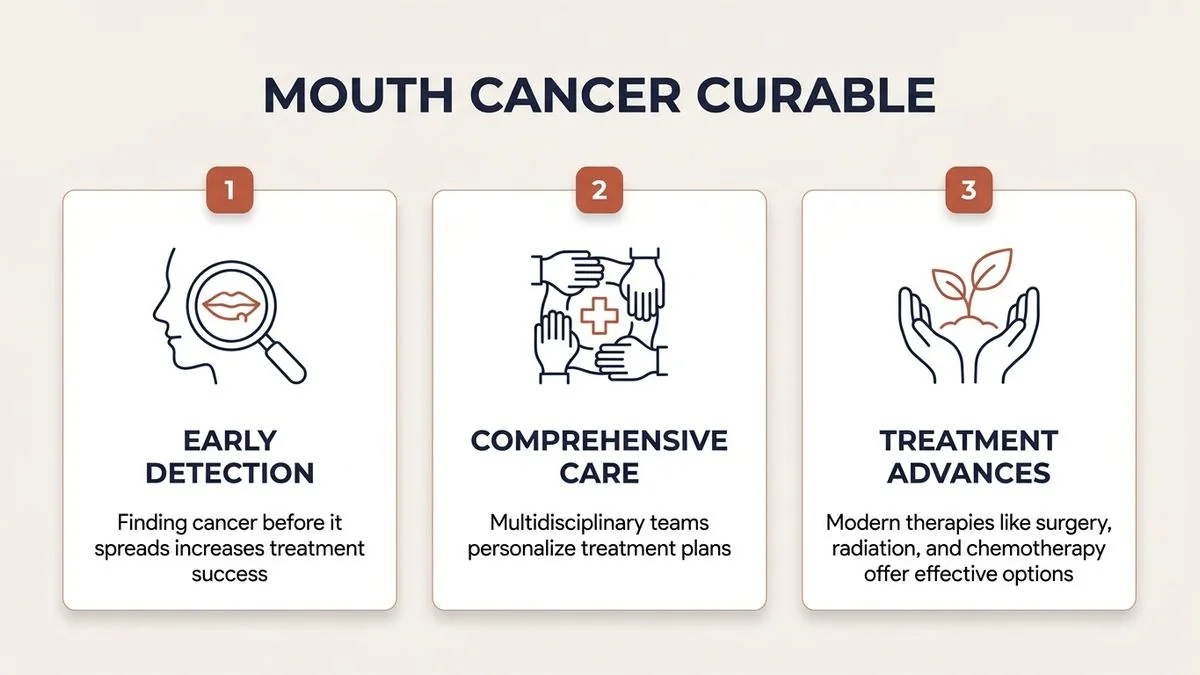
As oral and maxillofacial surgeons specializing in head and neck oncology, we address these vital concerns daily. In our Chennai practice, we emphasize that mouth cancer is highly treatable and, in many cases, completely curable. However, the path to a successful cure depends heavily on several clinical variables, with early detection and precise surgical execution serving as the cornerstones of a positive prognosis.
Understanding Mouth Cancer Curability: What Does "Cured" Mean?
In oncology, the term "cure" is often discussed in terms of long-term remission and survival milestones. For mouth cancer, clinicians typically evaluate treatment success using the 5-year relative survival rate. This statistic measures the percentage of patients who are alive five years after their initial diagnosis, compared to the general population.
If a patient remains free of cancer five years after completing their treatment, the likelihood of the disease returning drops significantly. At this stage, many clinicians consider the patient clinically cured.
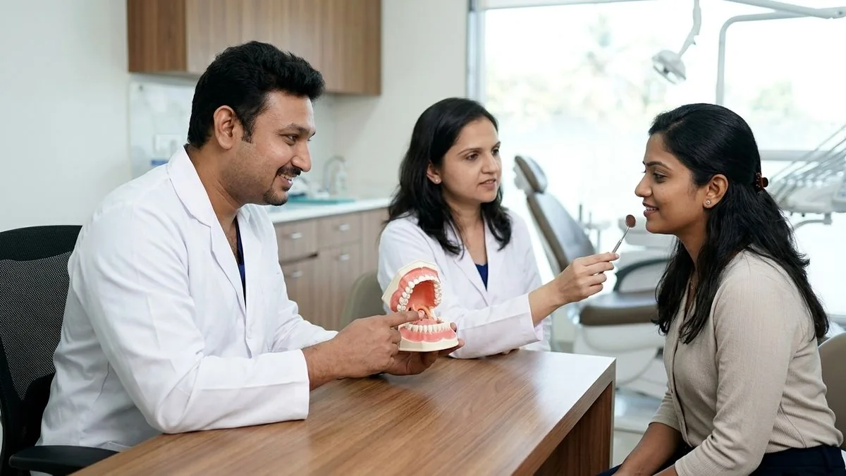
Achieving this milestone requires a highly coordinated treatment strategy. At our practice, Mouth Cancer Surgeons in Chennai, we utilize a unique dual-surgeon model. Both Dr. Pradeep S. and Dr. Kalpa Pandya personally evaluate, plan, and execute every procedure. This collaborative approach ensures that the oncological clearance of the tumor and the subsequent functional reconstruction of the oral cavity are planned in unison, directly optimizing both survival and post-surgical quality of life.
Key Factors That Influence Mouth Cancer Survival and Outlook
The prognosis for any individual diagnosed with oral cavity cancer is not determined by a single factor. Instead, it is shaped by a complex interplay of clinical, biological, and lifestyle elements.
1. The Stage at Diagnosis
The stage of the tumor at the time of initial diagnosis is the single most influential factor governing curability. Mouth cancer is staged using the TNM system:
- T (Tumor Size): How large is the primary tumor, and has it invaded deep structures like the jawbone, deep tongue muscles, or skin?
- N (Node Involvement): Has the cancer spread to the lymph nodes in the neck?
- M (Metastasis): Has the cancer spread to distant organs, such as the lungs or liver?
Cancers diagnosed at Stage I or II (localized, small tumors with no nodal spread) have exceptionally high cure rates. Conversely, Stage III and IV cancers (larger tumors that have spread to neck lymph nodes or distant sites) require more aggressive, multi-modality treatment and carry a more guarded prognosis.
2. Anatomical Location of the Tumor
The oral cavity contains several distinct sub-sites, each with its own clinical behavior. For example:
- Buccal Mucosa (Inner Cheek): Often associated with smokeless tobacco habits in South India. While highly treatable, these tumors can sometimes invade the deep facial spaces if left unchecked.
- Anterior Tongue: The tongue is highly vascular and rich in lymphatic drainage. Consequently, tongue cancers have a higher propensity to spread to the neck lymph nodes early, even when the primary tumor is relatively small. Understanding the types of oral cancer and their specific locations is vital for accurate prognostic planning.
- Floor of the Mouth: Tumors here can quickly involve the salivary glands and the lower jawbone (mandible), requiring meticulous surgical clearance. For a deeper look, read our guide on floor of mouth cancer.
3. Depth of Invasion (DOI)
In recent years, head and neck oncology guidelines (such as the AJCC 8th Edition) have placed immense clinical weight on the "Depth of Invasion" rather than just the surface size of the tumor. A thin, flat tumor that is 3 cm wide may actually have a better prognosis than a small, 1 cm tumor that has penetrated 10 mm deep into the muscle of the tongue. Deeply invasive tumors have a much higher risk of lymphatic metastasis, necessitating proactive treatment of the neck lymph nodes.
4. Surgical Margins
When performing oral cancer surgery, the primary objective is to achieve "clear margins." This means that when the pathologist examines the edges of the removed tissue under a microscope, no cancer cells are visible at the outer boundaries. Achieving negative (clear) margins is a primary predictor of local cure. If margins are positive or close (less than 1–2 mm), the risk of local recurrence increases, requiring additional surgery or postoperative radiation.
5. Patient Health and Habit Cessation
A patient’s general physical health plays a major role in how well they tolerate surgery and adjuvant therapies. Furthermore, immediate and permanent cessation of tobacco (smoking and chewing) and alcohol is mandatory. Continued use of these substances during and after treatment dramatically lowers survival rates and increases the risk of developing a second, entirely new primary cancer in the upper aerodigestive tract.
Survival Rates by Stage: What the Statistics Show
To understand the outlook of mouth cancer, it is helpful to examine established survival statistics. The data below is adapted from the National Cancer Institute's SEER (Surveillance, Epidemiology, and End Results) database, which categorizes oral cancers into localized, regional, and distant stages.
| Stage Category | Clinical Description | Estimated 5-Year Relative Survival Rate |
|---|---|---|
| Localized | The cancer is confined entirely to the primary site in the mouth (Stage I & II), with no spread to lymph nodes or other tissues. | 84% to 90% |
| Regional | The cancer has spread to nearby tissues or regional lymph nodes in the neck (Stage III & IV-A/B). | 65% to 68% |
| Distant | The cancer has metastasized to distant organs or structures outside the head and neck region (Stage IV-C). | 38% to 40% |
Note: These statistics are averages based on large populations. Individual outcomes can vary significantly based on the quality of surgical care, the genetic profile of the tumor, and the patient's response to therapy.
In our clinical experience in Chennai, patients who present early and undergo comprehensive, margin-free surgical resection frequently exceed these baseline statistical expectations, enjoying long, healthy, and highly functional lives.
The Role of Early Detection in Improving Prognosis
If there is one message we strive to convey to the public across Tamil Nadu and South India, it is this: early detection saves lives, saves jawbones, and preserves speech.
Many oral cancers do not begin as aggressive, deep-seated tumors. Instead, they often start as subtle, visible changes on the oral mucosa known as Oral Potentially Malignant Disorders (OPMDs). These include:
- Leukoplakia: Persistent white patches that cannot be scraped off.
- Erythroplakia: Smooth, red patches that carry a very high risk of dysplasia or early malignancy.
- Oral Submucous Fibrosis (OSMF): A chronic, scarring condition caused primarily by betel nut/areca nut chewing, characterized by progressive difficulty in opening the mouth.
[Healthy Oral Mucosa]
       │
       ▼
[OPMD / Precancerous Lesion (Leukoplakia/OSMF)]  <-- Best Window for Preventive Care!
       │
       ▼
[Early-Stage Mouth Cancer (Stage I/II)]        <-- High Cure Rate (~85%+), Simple Surgery
       │
       ▼
[Advanced-Stage Mouth Cancer (Stage III/IV)]    <-- Requires Complex Surgery, Flaps & Radiation
By identifying and treating these conditions early on through specialized OPMD care, we can often prevent oral cancer from developing entirely.
If a lesion has already transitioned into early-stage cancer, detecting it quickly means the surgical excision can be highly localized. This minimizes the impact on speech and swallowing, avoids the need for extensive bone removal, and frequently spares the patient from undergoing postoperative radiation therapy. If you have a persistent sore, read our resource on whether a mouth ulcer can be cancer or seek a professional oral cancer screening.
Mid-Article Guidance
If you are experiencing a non-healing mouth ulcer, a persistent white or red patch, or unexplained swelling in your mouth, do not wait. Early clinical evaluation is the most powerful tool to secure a favorable outcome.
Book a comprehensive consultation with Dr. Pradeep S. and Dr. Kalpa Pandya at Apollo Main Hospital, Greams Road, Chennai. Both surgeons review every case together to formulate a precise, personalized treatment plan.
Modern Surgical Interventions and Reconstruction
When a diagnosis of mouth cancer is confirmed, surgery is typically the primary treatment modality. The modern surgical management of oral cancer has evolved far beyond simply "removing the tumor." Today, it is a highly sophisticated discipline that balances complete oncological clearance with the meticulous restoration of oral form and function.
Wide Local Excision
The surgeon removes the primary tumor along with a safe three-dimensional margin of healthy tissue (usually 1 to 1.5 cm on all sides) to ensure no microscopic cancer cells are left behind.
Neck Dissection
Because oral cancers frequently spread to the lymph nodes in the neck, a neck dissection is often performed. During this procedure, the lymph nodes at risk are systematically removed and analyzed. In early-stage cancers with no clinical evidence of nodal spread, a "sentinel node biopsy" or a "selective neck dissection" may be performed as a preventive measure to eliminate micrometastases and accurately stage the disease.
Advanced Reconstructive Surgery
Removing a tumor from the tongue, cheek, or jaw leaves a structural defect. To restore the patient's ability to speak, swallow, and maintain their facial appearance, advanced reconstructive and restorative surgery is performed during the same operative session.
- Microvascular Free Flaps: This state-of-the-art technique involves transferring tissue (skin, muscle, or bone) from another part of the patient's body (such as the forearm, thigh, or fibula bone of the leg) and transplanting it to the oral cavity. The tiny blood vessels of the transferred tissue are micro-surgically sutured to blood vessels in the neck to restore blood flow.
- Composite Jaw Reconstruction: If a portion of the jawbone must be removed due to tumor invasion, we reconstruct it using a vascularized fibula free flap. This bone can later be fitted with specialized dental implants to fully restore chewing function.
Adjuvant Therapies: Boosting the Cure Rate
For advanced-stage mouth cancers (Stage III and IV), or when pathology reports reveal high-risk features (such as positive margins, perineural invasion, or spread to multiple lymph nodes), surgery alone may not be sufficient to guarantee a cure. In these cases, a multidisciplinary oncology approach is employed, combining surgery with adjuvant therapies:
- Radiation Therapy: High-energy X-rays are directed at the surgical site and the neck to destroy any remaining microscopic cancer cells.
- Chemotherapy: Medications are administered systemically to sensitize cancer cells to radiation or to target cells that may have escaped into the bloodstream.
- Targeted Therapy & Immunotherapy: Advanced molecular therapies that target specific pathways in cancer cells or stimulate the patient's own immune system to recognize and destroy tumor cells. These are increasingly utilized in recurrent or advanced cases to improve long-term survival.
By combining these modalities under the guidance of a multidisciplinary tumor board, we can significantly boost the cure rate and provide a positive outlook even for patients with advanced disease.
Post-Treatment Surveillance and Preventing Recurrence
The journey to being completely cured of mouth cancer does not end when the surgical wounds heal or the last session of radiation is completed. The first two years post-treatment represent the window of highest risk for local or regional recurrence.
A structured, rigorous surveillance protocol is essential to protect and maintain a patient's cancer-free status:
- Frequent Clinical Examinations: In our practice, we see patients every 4 to 6 weeks during the first year, gradually spacing out visits as time progresses.
- High-Resolution Imaging: Periodic ultrasounds of the neck, CT scans, or MRI scans are utilized to monitor deep tissue structures that cannot be fully evaluated by visual inspection alone.
- Endoscopy and Screening: Regular evaluations of the upper airway to screen for any secondary lesions.
- Lifestyle Maintenance: Continuous support for habit cessation, nutritional optimization, and oral hygiene management.
The core advantage of our dual-surgeon model at Mouth Cancer Surgeons, Chennai, is continuity of care. The same two surgeons—Dr. Pradeep S. and Dr. Kalpa Pandya—who perform your initial biopsy and execute your complex surgery will be the exact same specialists conducting your long-term follow-up examinations. This deep, longitudinal familiarity with your specific surgical anatomy is invaluable for identifying subtle, early changes that might indicate a recurrence.
Patient-Centric Perspective: Living Beyond Mouth Cancer
Survival is about more than just clinical cure rates; it is about reclaiming your quality of life. Modern oral oncology places a profound emphasis on functional rehabilitation.
Following surgery and reconstructive procedures, patients often work closely with a dedicated rehabilitation team:
- Speech and Swallowing Therapy: Specialized exercises to help patients adapt to changes in the tongue or palate, ensuring they can speak clearly and eat safely.
- Dietary and Nutritional Support: Tailored nutritional plans to maintain weight and strength during and after treatment.
- Pre-Prosthetic and Dental Implant Rehabilitation: Once the reconstructed jaw has fully healed and the patient is cleared of active disease, we can place specialized dental implants into the reconstructed bone. This allows for the fabrication of fixed dental prostheses, restoring the natural smile and the ability to chew solid foods.
While a diagnosis of mouth cancer is undeniably life-altering, the combination of early detection, advanced microvascular surgery, and dedicated rehabilitation means that patients can look forward to a future that is not only cancer-free but also rich in function, dignity, and comfort.
Consult Mouth Cancer Surgeons at Apollo Main Hospital, Chennai
If you or a loved one are navigating a diagnosis of oral cancer, seeking a second opinion, or experiencing concerning symptoms like non-healing ulcers or difficulty opening your mouth, expert guidance is available.
At Mouth Cancer Surgeons, Chennai, Dr. Pradeep S. (MDS, FHNO, FIBCSOMS) and Dr. Kalpa Pandya (MDS, FHNS) bring decades of combined experience, having treated thousands of patients across South India. By combining world-class surgical oncology with state-of-the-art microvascular reconstruction, they work side-by-side to deliver the highest standard of personalized care.
Take the first step toward clarity and healing today:
- Primary Consultation Location: Apollo Main Hospital, Greams Road, Thousand Lights, Chennai.
- Direct Enquiry / Appointment Booking: +91 96633 03747
- Online Booking: Request an Appointment
References
- National Cancer Institute. "Cancer Stat Facts: Oral Cavity and Pharyngeal Cancer." SEER Program, 2023. https://seer.cancer.gov/statfacts/html/oralcav.html
- Amin, M. B., et al. "AJCC Cancer Staging Manual (8th Edition) - Head and Neck Tumors." Springer, 2017.
- National Comprehensive Cancer Network (NCCN). "NCCN Clinical Practice Guidelines in Oncology: Head and Neck Cancers." NCCN Guidelines, 2024. https://www.nccn.org/guidelines
- Indian Council of Medical Research (ICMR). "Consensus Document for the Management of Buccal Mucosa Cancer." ICMR Guidelines, 2020.
- Shah, J. P., Patel, S. G., & Singh, B. "Head and Neck Surgery and Oncology." Elsevier Health Sciences, 2019.
Authored by
Medically reviewed by