Floor of Mouth Cancer Symptoms and Warning Signs to Note
Need expert consultation? Book an appointment with Dr. Pradeep S. or Dr. Kalpa Pandya.
Book AppointmentThe floor of the mouth is a small but highly complex anatomical zone. Located beneath the tongue and bounded by the lower jawbone (mandible), this horseshoe-shaped region is lined with a delicate mucosal membrane. Because of its hidden location, changes in this area are frequently overlooked during routine daily hygiene.
Recognizing floor of mouth cancer symptoms and warning signs early can make a profound difference in treatment outcomes. This guide explains what to look for, how to distinguish benign changes from malignancy, and when to seek professional evaluation from specialist oral and maxillofacial surgeons.
Understanding the Anatomy of the Floor of the Mouth
To understand how symptoms develop, it helps to understand the anatomy of the sublingual space. The floor of the mouth contains several vital structures:
- The Sublingual and Submandibular Glands: Salivary glands that release saliva through tiny ducts (including Wharton's duct) under the tongue.
- The Lingual Nerve: Provides sensation to the anterior two-thirds of the tongue and the floor of the mouth.
- The Hypoglossal Nerve: Controls the movement of the tongue muscles.
- Mylohyoid and Genioglossus Muscles: Structural muscles that form the muscular floor of the mouth and control tongue protrusion and swallowing.
Because this region is highly vascular and rich in lymphatic drainage channels, cancers that develop here can grow quickly and spread to the lymph nodes in the neck (cervical lymph nodes) if not diagnosed early. More than 90% of malignant tumors in this area are oral squamous cell carcinomas (OSCC), arising from the flat squamous cells that line the mucosal surface.
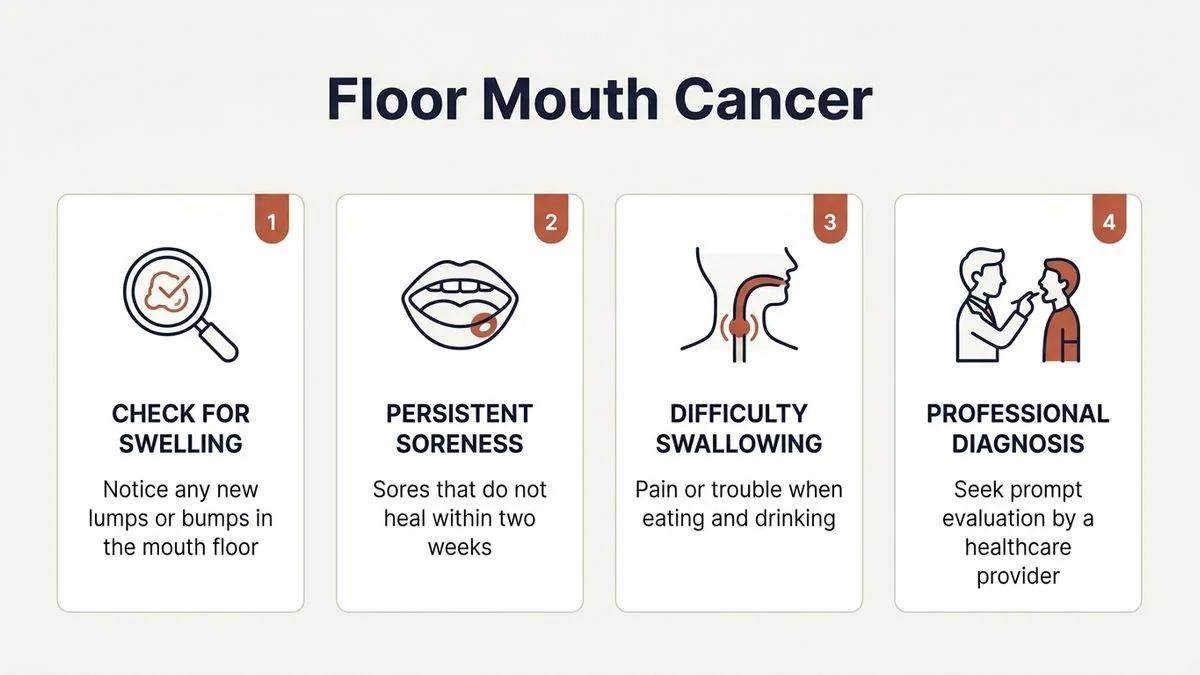
Early Floor of Mouth Cancer Symptoms and Warning Signs
In its earliest stages, floor of mouth cancer is often completely silent and painless. This lack of pain is the primary reason many patients delay seeking medical attention.
1. Persistent Red or White Patches (Erythroplakia and Leukoplakia)
One of the earliest warning signs is a change in the color and texture of the mucosal lining under the tongue.
- Leukoplakia: These are flat, slightly raised white patches that cannot be scraped off with a toothbrush or gauze. While some leukoplakias are benign, others represent oral precancer (OPMD) or early invasive cancer.
- Erythroplakia: These are bright red, velvety patches. Erythroplakia carries a significantly higher risk of dysplasia (precancerous changes) or malignancy than white patches.
- Erythroleukoplakia: A mixed red-and-white patch that is highly suspicious and warrants immediate biopsy.
2. A Non-Healing Ulcer under the Tongue
A classic mouth ulcer warning sign is a sore that does not heal. While common canker sores or traumatic ulcers (from a sharp tooth or ill-fitting denture) typically heal within 10 to 14 days, a cancerous ulcer persists.
- Characteristics of a cancerous ulcer: It often has raised, rolled, or firm edges (induration). The center of the ulcer may look raw, granular, or bleed easily when touched.
3. A Painless Lump or Thickening
You may feel a small, firm lump or an area of localized thickening in the tissue under your tongue using your tongue or finger. In our clinical practice, patients often describe feeling a "fleshy tag" or a "hard spot" that slowly grows larger over several weeks or months.
Advanced Symptoms: When the Disease Progresses
If early signs are ignored, the tumor can invade the deeper muscular, neural, and bony structures of the oral cavity. This deep invasion produces more pronounced, functional symptoms.
1. Unexplained Pain and Tenderness
As the tumor grows deeper, it eventually irritates sensory nerve endings, causing a persistent, localized ache or a sharp pain under the tongue. This pain may worsen during chewing, swallowing, or speaking.
2. Referred Otalgia (Ear Pain)
A highly specific but frequently misunderstood symptom of advanced floor of mouth cancer is referred ear pain.
- The Neural Pathway: The lingual nerve, which provides sensation to the floor of the mouth, travels close to the mandibular branch of the trigeminal nerve. When a tumor invades deep tissues, pain signals travel along these shared neural pathways.
- The Result: The brain misinterprets the pain as coming from the ear on the same side as the mouth lesion, even though the ear itself is completely healthy. If you have persistent ear pain alongside a sore under your tongue, it requires an urgent oral oncology evaluation.
3. Restricted Tongue Movement (Ankyloglossia)
The tongue relies on a complex network of muscles to move freely for speech and swallowing. When a floor of mouth tumor invades the underlying genioglossus or hyoglossus muscles, it anchors the tongue to the floor of the mouth.
- What patients notice: Difficulty protruding the tongue, slurred speech (dysarthria), or a feeling that the tongue is "stuck" or heavy.
4. Difficulty Swallowing (Dysphagia) and Painful Swallowing (Odynophagia)
As the tumor expands, it physically obstructs the passage of food and saliva. The coordinated muscular movements required for safe swallowing become painful and inefficient, leading to choking, coughing, or a sensation of food being stuck in the throat.
5. Loose Teeth or Jaw Pain
If the tumor spreads laterally, it can invade the periosteum (the outer membrane) and the cortex of the mandible (lower jawbone). This can lead to:
- Unexplained loosening of the lower front teeth or premolars.
- Deep, dull aching pain in the jawbone.
- Numbness in the lower lip or chin (caused by involvement of the inferior alveolar nerve).
6. A Lump in the Neck
Often, the first symptom that prompts a patient to visit a doctor is a painless, firm swelling in the neck. This occurs when cancer cells travel through the lymphatic vessels to the submental or submandibular lymph nodes (located just under the chin and jawline). A neck lump that is hard, fixed to surrounding tissues, and gradually enlarging is a strong indicator of regional metastasis.
Benign Ulcers vs. Floor of Mouth Cancer: Key Differences
It is common to experience minor mouth sores, and not every lesion under the tongue is cancerous. The table below highlights the key clinical differences between benign conditions and potential malignancies:
| Clinical Feature | Benign Oral Lesion (e.g., Aphthous Ulcer, Trauma) | Floor of Mouth Cancer (Malignancy) |
|---|---|---|
| Duration | Heals completely within 10 to 14 days. | Persists for more than 2 to 3 weeks and continues to grow. |
| Pain | Highly painful at onset, gradually improving. | Often painless in early stages; deep, dull ache in later stages. |
| Texture & Margins | Soft, flexible borders; flat or slightly depressed center. | Firm, hard to the touch (indurated); raised, rolled, or irregular borders. |
| Bleeding | Rarely bleeds unless directly irritated. | Bleeds easily upon minor contact or bimanual palpation. |
| Associated Patches | No surrounding red or white changes. | Often surrounded by leukoplakia (white) or erythroplakia (red) patches. |
| Neck Lymph Nodes | May be tender and soft (reactive) if infected; resolve quickly. | Hard, painless, fixed, and progressively enlarging neck swellings. |
High-Risk Factors and Etiology
Understanding your risk profile is an essential part of evaluating symptoms. While anyone can develop floor of mouth cancer, certain lifestyle factors significantly increase the risk:
- Tobacco Use: Smoking cigarettes, bidis, or cigars, as well as using smokeless tobacco (gutka, khaini, mawa), introduces potent carcinogens directly to the mucosal lining. The floor of the mouth acts as a "sump" or reservoir where tobacco juices pool, leading to prolonged exposure.
- Alcohol Consumption: Alcohol acts as a solvent, dehydrating the mucosal lining and making it easier for carcinogens to penetrate the cells.
- The Synergistic Effect: Individuals who both smoke and drink alcohol have a risk of developing oral cancer that is up to 15 times higher than those who do neither.
- Betel Quid/Areca Nut Chewing: Extremely common across Tamil Nadu and South India, chewing betel nut causes chronic mechanical and chemical irritation, often leading to oral submucous fibrosis (OSMF), a high-risk precancerous condition.
- Poor Oral Hygiene and Chronic Irritation: Sharp, broken teeth or ill-fitting dentures that rub continuously against the floor of the mouth can cause chronic inflammation, which may contribute to malignant transformation over time.
How Floor of Mouth Cancer is Diagnosed
If you present with suspicious symptoms, our team at Mouth Cancer Surgeons follows a systematic diagnostic protocol to ensure an accurate and rapid assessment.
1. Comprehensive Clinical Examination & Bimanual Palpation
The first step is a thorough visual and physical examination. Because tumors in the floor of the mouth can grow downward into the deeper soft tissues without showing significant surface changes, bimanual palpation is essential.
During this exam, the surgeon places one gloved finger inside your mouth under the tongue and the fingers of the other hand on the outside of your neck under your jaw. By gently pressing the tissues together, the surgeon can feel for deep-seated hardness, lumps, or structural thickening that might not be visible to the naked eye.
2. Tissue Biopsy
A biopsy is the only definitive way to diagnose oral cancer.
- Incisional Biopsy: Under local anesthesia, a small sample of tissue is taken from the edge of the ulcer or lump. This procedure is quick, virtually painless, and performed in an outpatient setting.
- Histopathology: The tissue is sent to a specialized oral pathologist who examines the cells under a microscope to determine if they are cancerous and, if so, to identify the specific type of oral cancer.
3. Imaging Studies
If the biopsy confirms oral squamous cell carcinoma, imaging is performed to evaluate the extent of the tumor:
- Contrast-Enhanced CT (CECT) Scan: Helps evaluate if the tumor has invaded the cortical bone of the mandible (lower jaw) and assesses the neck lymph nodes for metastasis.
- Magnetic Resonance Imaging (MRI): Provides superior soft-tissue contrast, allowing the surgeon to see exactly how deeply the tumor has invaded the tongue muscles and adjacent sublingual spaces.
- Orthopantomogram (OPG): A panoramic dental X-ray used to check the general health of the teeth and jawbone.
Early Detection Saves Lives: The Role of Screening
Like most head and neck cancers, the stage at diagnosis is the single most important factor determining the success of treatment.
According to global oncology data, when floor of mouth cancer is detected at an early stage (Stage I or II), the five-year survival rate is excellent, often exceeding 80% to 90%. Conversely, advanced-stage cancers (Stage III or IV) require more extensive surgery, radiation, and chemotherapy, and have lower long-term survival rates.
Regular oral cancer screening is simple, non-invasive, and takes less than ten minutes. If you are a high-risk individual (tobacco or alcohol user) or have noticed any persistent changes in your mouth, scheduling a screening can help catch issues before they progress.
If you are experiencing any of these warning signs, early consultation is important. Book an appointment with Dr. Pradeep S. and Dr. Kalpa Pandya at Apollo Main Hospital, Greams Road, Chennai.
Surgical and Reconstructive Considerations
If a diagnosis of floor of mouth cancer is confirmed, surgery is typically the primary treatment modality. The goal of surgery is to remove the tumor completely with safe, cancer-free margins while preserving as much oral function as possible.
1. Tumor Resection
The extent of the surgery depends on the tumor's size and depth:
- Wide Local Excision: For small, superficial tumors, the lesion is removed along with a surrounding margin of healthy tissue.
- Marginal or Segmental Mandibulectomy: If the cancer has grown close to or invaded the jawbone, a portion of the lower jaw must be removed to ensure complete clearance.
- Neck Dissection: Because floor of mouth cancers have a high rate of early spread to the neck, the lymph nodes under the jaw and along the side of the neck are often removed (either as a preventive measure or to treat known disease).
2. Reconstructive & Restorative Surgery
The floor of the mouth plays a vital role in speech, swallowing, and keeping saliva inside the oral cavity. Removing tissue from this area can impact these functions if not reconstructed properly.
At Mouth Cancer Surgeons, our dual-surgeon team specializes in advanced reconstructive and restorative surgery. Using microvascular free flaps (such as tissue taken from the forearm or thigh), we can reconstruct the floor of the mouth, rebuild the jawbone if necessary, and restore both appearance and oral function.
The Dual-Surgeon Advantage: Dr. Pradeep S. & Dr. Kalpa Pandya
Navigating a potential cancer diagnosis can be overwhelming. At Mouth Cancer Surgeons, we believe in a highly personalized, continuous model of care.
Our practice is led by two dedicated oral and maxillofacial surgeons who manage your care together from your initial consultation and diagnostic biopsy, through complex surgery and reconstruction, to long-term follow-up:
- Dr. Pradeep S. (MDS, FHNO, FIBCSOMS, PGDCR) is an internationally board-certified consultant oral and maxillofacial surgeon with over 7 years of specialized experience in head and neck surgical oncology. He leads our advanced oral cancer resections and complex microvascular reconstructions. He consults at Apollo Main Hospital, Greams Road, Chennai.
- Dr. Kalpa Pandya (BDS, MDS, FHNS) is a consultant oral and maxillofacial surgeon with over 10 years of experience who has cared for more than 1,000 oral cancer patients. She leads our clinical screenings, management of oral precancers (OPMDs), facial trauma, and dental implant rehabilitation. She is also an Assistant Professor and Associate Consultant at Sri Ramachandra Medical Centre, Porur, Chennai.
By combining our expertise, we ensure that every patient benefits from two specialist opinions on every case, resulting in precise surgical planning, meticulous reconstruction, and compassionate supportive care.
Summary Checklist: When to Consult a Specialist
If you or a loved one notices any of the following symptoms, do not wait for them to resolve on their own. Schedule a clinical examination if you experience:
- A red, white, or mixed patch under the tongue that persists for more than two weeks.
- An ulcer, sore, or raw area in the floor of the mouth that does not heal within 14 days.
- A firm lump, hard spot, or localized thickening under the tongue.
- Persistent ear pain on one side with no obvious ear infection.
- Difficulty moving your tongue freely, slurred speech, or trouble swallowing.
- A painless, firm swelling or lump in your neck or under your jawline.
- Unexplained loose teeth in the lower jaw or persistent numbness in your lower lip.
Have questions about your condition? Request a consultation with our oral & maxillofacial surgeons — both surgeons review every case together.
For personalised treatment options and expert care, consult Dr. Pradeep S. and Dr. Kalpa Pandya — Mouth Cancer Surgeons, Chennai. Call +91 96633 03747 or book an appointment.
References
- National Comprehensive Cancer Network (NCCN). "Clinical Practice Guidelines in Oncology: Head and Neck Cancers." NCCN Guidelines, 2025. https://www.nccn.org
- World Health Organization. "Oral Cancer Prevention and Control." WHO Technical Report Series, 2024. https://www.who.int
- Shah, Jatin P., et al. Head and Neck Surgery and Oncology. 5th ed., Elsevier, 2020.
- Indian Council of Medical Research (ICMR). "Consensus Document for Management of Buccal Mucosa and Oral Cavity Cancers." ICMR Guidelines, 2022.
- National Cancer Institute. "Lip and Oral Cavity Cancer Treatment (PDQ®)–Health Professional Version." National Institutes of Health, 2025. https://www.cancer.gov
- Lydiatt, D. D., et al. "AJCC Cancer Staging Manual: Head and Neck Sites." CA: A Cancer Journal for Clinicians, 2017. https://acsjournals.onlinelibrary.wiley.com
Authored by

Medically reviewed by