Types of Oral Cancer: An Overview for Chennai Patients
Need expert consultation? Book an appointment with Dr. Pradeep S. or Dr. Kalpa Pandya.
Book AppointmentWhen patients hear the term "mouth cancer," they often assume it refers to a single, uniform disease. In reality, the oral cavity is home to many different tissue types, including surface membranes, salivary glands, muscle, bone, and blood vessels. Malignant changes can occur in any of these tissues, resulting in several distinct types of oral cancer.
Understanding the specific type of oral cancer is the first and most critical step in planning treatment. The cellular characteristics of a tumor dictate how rapidly it may grow, how likely it is to spread to the lymph nodes in the neck, and which surgical or non-surgical treatments will be most effective.
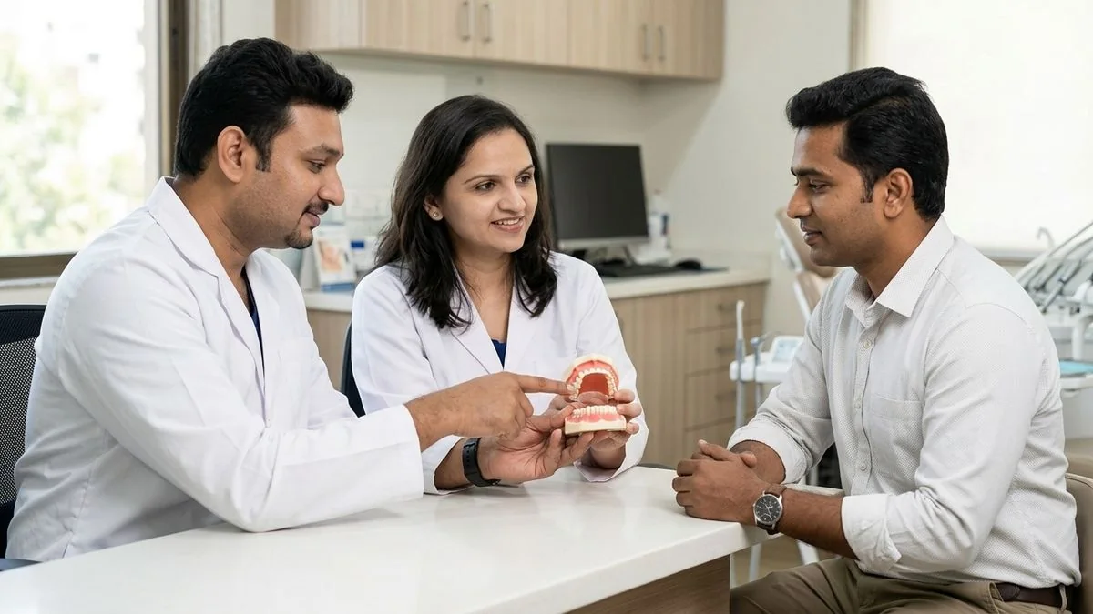
At Mouth Cancer Surgeons in Chennai, our dual-surgeon team—Dr. Pradeep S. and Dr. Kalpa Pandya—collaborates on every case. From the initial clinical examination and diagnostic biopsy at Apollo Main Hospital, Greams Road, to the execution of complex oncological resections and advanced microvascular reconstructions, we ensure our patients benefit from our combined expertise. This comprehensive overview of the types of oral cancer is designed to help patients, families, and caregivers understand the pathological landscape of oral malignancies.
Understanding the Oral Cavity: Where Malignancies Form
To understand the different types of oral cancer, it helps to look at the anatomy of the oral cavity. The oral cavity begins at the border of the lips and extends back to the junction where the hard palate meets the soft palate.
The primary anatomical sites within the oral cavity include:
- The Lips: Particularly the mucosal border where the lip meets the skin (vermilion border).
- The Oral Tongue: The highly mobile anterior two-thirds of the tongue.
- The Buccal Mucosa: The inner lining of the cheeks.
- The Floor of the Mouth: The horseshoe-shaped area under the tongue.
- The Alveolar Ridge and Gingiva: The gums surrounding the upper and lower teeth.
- The Hard Palate: The bony roof of the mouth.
- The Retromolar Trigone: The small area of mucosa behind the lower wisdom teeth.
Each of these areas is lined by a protective layer of flat, scale-like cells called squamous cells. Beneath this lining lie minor salivary glands, connective tissues, nerves, blood vessels, and the jawbones (the maxilla and mandible). Because different types of cells populate these layers, several distinct forms of cancer can arise here.
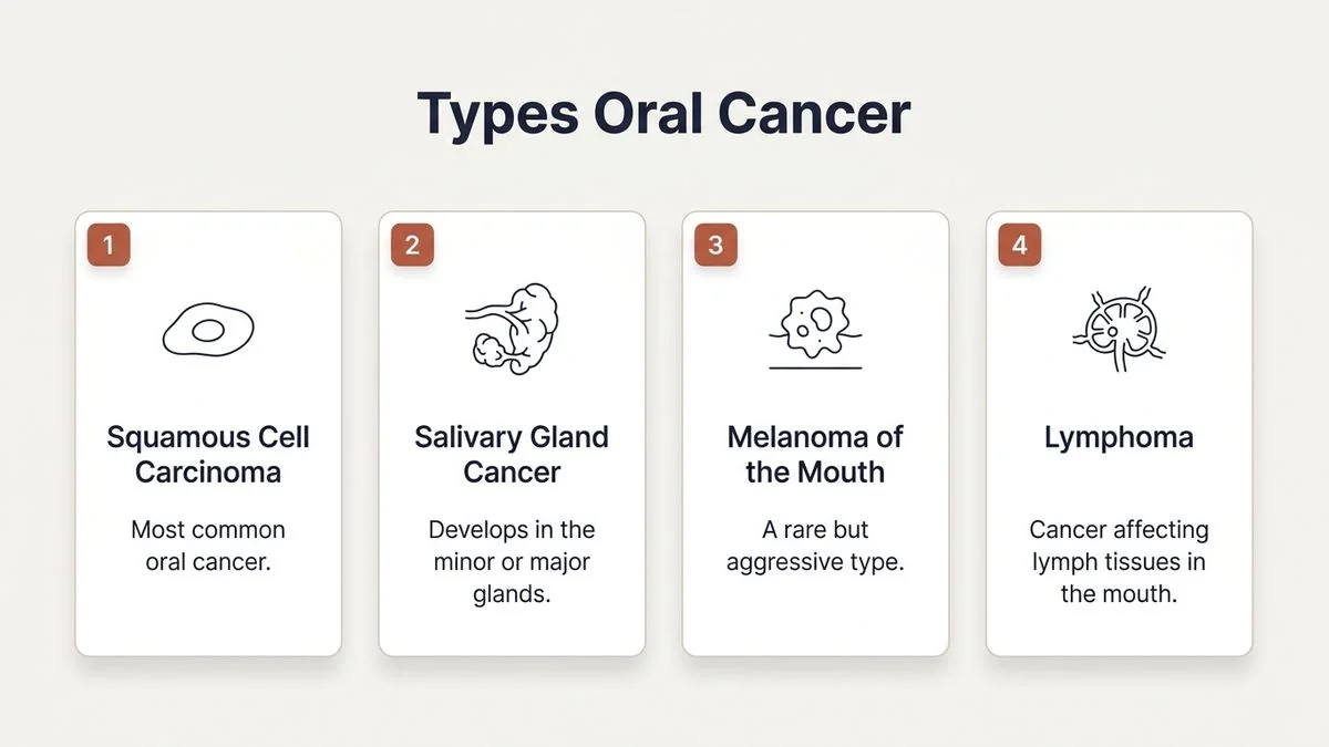
Squamous Cell Carcinoma (SCC): The Most Common Oral Cancer
Squamous Cell Carcinoma (SCC) is the most common type of oral cancer, accounting for more than 90% of all malignancies diagnosed in the oral cavity. This cancer begins in the squamous cells that form the thin, protective surface layer (epithelium) of the oral mucosa.
Over time, chronic irritation from risk factors such as tobacco use (both smoking and smokeless forms like gutka or betel quid), heavy alcohol consumption, and nutritional deficiencies can cause genetic mutations in these cells. These mutations lead to uncontrolled cell division, eventually breaking through the basement membrane of the epithelium to invade deeper tissue layers.
Subtypes and Variants of Oral SCC
Not all squamous cell carcinomas behave the same way. Pathologists classify SCC into several variants based on how the cells look under a microscope:
- Well-Differentiated SCC: The cancer cells closely resemble normal squamous cells. They tend to grow more slowly and are less aggressive.
- Moderately Differentiated SCC: The cells show intermediate abnormalities. This is the most common presentation and requires decisive surgical intervention.
- Poorly Differentiated or Anaplastic SCC: The cells look highly abnormal and disorganized. This variant is aggressive, grows rapidly, and carries a higher risk of spreading (metastasizing) to the cervical lymph nodes in the neck.
- Verrucous Carcinoma: This is a distinct, highly differentiated variant of SCC. It often presents as a slow-growing, wart-like, white patch. While it can become quite large and locally destructive to surrounding bone and soft tissue, it rarely metastasizes to lymph nodes. It is highly associated with the use of smokeless tobacco.
- Spindle Cell Carcinoma (Sarcomatoid Carcinoma): A rare and highly aggressive variant of SCC where the cells take on a spindle-like shape, resembling a sarcoma. It requires prompt, wide surgical excision.
In our practice, we emphasize that early-stage SCC can mimic benign oral conditions. A persistent, painless red or white patch or an ulcer that does not heal within two weeks warrants immediate professional evaluation. You can read more about identifying these early changes in our article on can a mouth ulcer be cancer.
Salivary Gland Malignancies: Not All Mouth Cancers Are Squamous
While the major salivary glands (parotid, submandibular, and sublingual) sit outside the oral cavity proper, hundreds of microscopic minor salivary glands are scattered throughout the lining of the mouth. These glands are located primarily in the hard palate, the inner cheeks, and the lips.
Malignancies arising from these minor salivary glands account for approximately 3% to 5% of oral cavity cancers. Unlike SCC, which begins on the surface lining, minor salivary gland cancers develop deep within the gland tissue, often presenting as a firm, painless, submucosal lump covered by normal-looking mucosa.
The most common malignant minor salivary gland tumors include:
Mucoepidermoid Carcinoma
This is the most common salivary gland malignancy. It contains a mix of mucus-producing cells and squamous-like epithelial cells. They are graded as low, intermediate, or high grade. Low-grade mucoepidermoid carcinomas are slow-growing and have an excellent prognosis with conservative surgical removal, whereas high-grade tumors behave aggressively.
Adenoid Cystic Carcinoma (ACC)
Adenoid cystic carcinoma is characterized by its unique ability to grow along nerve pathways, a feature known as perineural invasion. This can cause localized pain, numbness, or weakness in the facial muscles. ACC is known for its slow but relentless growth pattern and a tendency to recur locally or spread to distant sites like the lungs, even years after successful primary treatment.
Adenocarcinoma
This is a broad category of salivary gland cancers that develop in the glandular ductal cells. Like other salivary malignancies, they require wide surgical excision, often followed by radiation therapy to ensure local control.
Because minor salivary gland tumors often present as painless lumps beneath intact skin or mucosa, they are frequently misdiagnosed as benign cysts or dental infections in their early stages. A high index of clinical suspicion and advanced imaging are crucial for accurate identification.
Rare Types of Oral Cancer: Sarcomas, Melanomas, and Lymphomas
While squamous cell carcinoma and salivary gland tumors make up the vast majority of oral malignancies, other rare cancers can develop from the deeper structural tissues of the mouth.
1. Oral Mucosal Melanoma
Melanoma is most commonly associated with the skin, but it can also develop in the moist mucosal linings of the body. Oral mucosal melanoma is an extremely rare and highly aggressive cancer, representing less than 1% of all oral malignancies.
- Presentation: It typically presents as a dark brown, black, or occasionally pigment-free (amelanotic) patch or lump, most commonly on the hard palate or upper gums.
- Behavior: It has a strong tendency to invade deep tissue early and spread rapidly through the bloodstream and lymphatic system. Wide surgical resection combined with immunotherapy or targeted therapy is usually required.
2. Maxillofacial Sarcomas
Sarcomas are cancers that arise from connective tissues such as bone, cartilage, fat, muscle, or blood vessels. In the oral cavity, they can develop within the upper jaw (maxilla) or lower jaw (mandible).
- Osteosarcoma of the Jaw: A malignant bone tumor that presents as a rapidly expanding swelling of the jaw, loose teeth, or unexplained facial pain.
- Chondrosarcoma: A rare malignant tumor that produces cartilage.
- Fibrosarcoma and Rhabdomyosarcoma: Malignancies of the fibrous connective tissue and skeletal muscle, respectively.
Patients with jaw swelling, shifting teeth, or unexplained numbness in the chin or lip require comprehensive clinical and radiological evaluation to rule out these bone and soft tissue pathologies. Specialized management for these conditions is detailed on our maxillofacial tumours & jaw pathology page.
3. Oral Lymphomas
Lymphomas are cancers of the lymphatic system. While they typically start in the lymph nodes, they can occasionally develop in extranodal sites, including the oral cavity. The tonsils, base of the tongue, and palate are the most common sites. Oral lymphomas often present as soft, fleshy, non-ulcerated swellings and are generally treated with chemotherapy and radiation rather than primary surgery.
Oral Potentially Malignant Disorders (OPMD): Pre-Cancerous Changes
Many oral squamous cell carcinomas do not appear suddenly; they develop from pre-existing, non-cancerous tissue changes known as Oral Potentially Malignant Disorders (OPMD). Recognizing and treating these conditions early can prevent them from transforming into invasive cancer.
Dr. Kalpa Pandya leads our practice's focus on OPMD management, helping patients manage high-risk lesions through systematic monitoring, medical therapy, or conservative surgical excision.
| OPMD Type | Clinical Appearance | Risk of Malignant Transformation | Primary Causes / Associations |
|---|---|---|---|
| Leukoplakia | Persistent white patch that cannot be scraped off or attributed to other physical trauma. | Variable (1% to 20%), higher if it shows dysplasia on biopsy. | Smoking, smokeless tobacco, chronic friction. |
| Erythroplakia | Smooth, velvety red patch that is often flat or slightly depressed. | Very High (over 50% demonstrate dysplasia or early cancer on biopsy). | Tobacco and alcohol abuse. |
| Oral Submucous Fibrosis (OSMF) | Progressive scarring and stiffening of the oral mucosa, leading to restricted mouth opening (trismus). | Moderate to High (approximately 7% to 13%). | Areca nut (betel nut) chewing, highly prevalent in South India. |
| Erythroleukoplakia | A mixed red-and-white lesion, often with a speckled or nodular surface. | High risk of harboring invasive cancer. | Combined tobacco and alcohol use. |
If you have been diagnosed with any of these lesions, regular follow-ups and tissue biopsies are essential to catch cellular changes (dysplasia) before they progress to invasive cancer. Learn more about our preventive care on the oral precancer (OPMD) page.
How Oral Cancers Are Staged and Graded
Once a biopsy confirms the specific type of oral cancer, our team determines the stage of the disease. Staging describes how far the cancer has spread within the oral cavity and whether it has reached nearby lymph nodes or other organs.
We utilize the globally recognized TNM Staging System established by the American Joint Committee on Cancer (AJCC):
- T (Tumor Size and Depth of Invasion): Measures the size of the primary tumor and how deeply it has grown into the underlying tissues. Depth of invasion (DOI) is a critical factor in oral tongue and floor of mouth cancers, as deeper tumors have a much higher likelihood of spreading to the neck.
- N (Node Involvement): Evaluates whether the cancer has spread to the lymph nodes in the neck, including the number, size, and location of the affected nodes.
- M (Metastasis): Identifies whether the cancer has spread to distant parts of the body, such as the lungs, liver, or bones.
The Stages of Oral Cancer
- Stage I: The tumor is small (2 cm or less) and superficial, with no spread to lymph nodes.
- Stage II: The tumor is larger (between 2 and 4 cm) but remains localized without lymph node involvement.
- Stage III: The tumor is larger than 4 cm, or has invaded deeper tissues, or has spread to a single lymph node on the same side of the neck.
- Stage IV: Advanced disease. This includes tumors that have invaded nearby structures (like the jawbone, deep tongue muscles, or skin of the face), spread to multiple lymph nodes, or metastasized to distant organs.
Understanding the stage is vital because it determines the complexity of the treatment plan. While early-stage cancers (Stages I and II) can often be treated with a single modality—typically surgical resection—advanced stages (Stages III and IV) require a comprehensive approach combining surgery, microvascular reconstruction, radiation therapy, and chemotherapy. To understand more about treatment success and prognosis based on these stages, read our detailed guide on is mouth cancer curable.
Comprehensive Comparison of Oral Cavity Malignancies
To help patients visualize the differences between the major types of oral cavity cancers, the following table summarizes their clinical characteristics, common locations, and primary treatment approaches.
| Cancer Type | Primary Originating Cells | Common Locations | Growth Pattern & Behavior | Primary Treatment Approach |
|---|---|---|---|---|
| Squamous Cell Carcinoma (SCC) | Surface epithelial squamous cells | Tongue, floor of mouth, buccal mucosa, lower lip | Can be rapid; frequently spreads to neck lymph nodes | Wide surgical excision + neck dissection, followed by reconstruction. Adjuvant radiation/chemotherapy for advanced stages. |
| Verrucous Carcinoma | Well-differentiated squamous cells | Buccal mucosa, gingiva | Slow-growing, outward-pushing, locally destructive; rarely spreads to lymph nodes | Wide surgical excision. Radiation is generally avoided due to risk of anaplastic transformation. |
| Mucoepidermoid Carcinoma | Minor salivary gland ductal cells | Hard palate, buccal mucosa | Variable; low-grade is slow and localized, high-grade is aggressive | Surgical excision with clear margins. Radiation therapy is added for high-grade or close margins. |
| Adenoid Cystic Carcinoma | Minor salivary gland cells | Hard palate, upper lip | Slow but highly infiltrative; notable for growing along nerves (perineural invasion) | Wide surgical resection, almost always followed by post-operative radiation therapy due to perineural spread. |
| Oral Mucosal Melanoma | Melanocytes (pigment cells) | Hard palate, maxillary gingiva | Extremely aggressive; early deep invasion and distant metastasis | Radical surgical resection combined with systemic immunotherapies or targeted therapies. |
| Osteosarcoma of the Jaw | Bone-forming osteoblasts | Mandible (lower jaw), Maxilla (upper jaw) | Rapidly expanding bone swelling, pain, loose teeth | Neoadjuvant chemotherapy followed by radical surgical resection (jaw resection) and microvascular bone reconstruction. |
The Dual-Surgeon Model: Why Collaborative Care Matters
At Mouth Cancer Surgeons, we believe that treating oral cancer requires a highly coordinated, multidisciplinary approach. The anatomy of the head and neck is incredibly complex, containing vital structures responsible for breathing, swallowing, speech, and facial appearance.
Our practice operates on a unique dual-surgeon model, where Dr. Pradeep S. and Dr. Kalpa Pandya evaluate, plan, and operate together on complex cases.
- Surgical Precision: During major surgeries, having two highly trained oral and maxillofacial surgeons working in tandem reduces anesthesia time and increases surgical safety. While one surgeon focuses on the precise oncological clearance of the tumor and neck dissection, the other prepares the reconstructive graft.
- Advanced Reconstruction: Restoring function and appearance is as important as removing the cancer. Dr. Pradeep S. specializes in head and neck surgical oncology and microvascular free flap reconstructions (using tissue from the forearm, fibula, or thigh to rebuild the tongue or jawbone). This allows patients to regain the ability to speak, chew, and swallow.
- Comprehensive Rehabilitation: Dr. Kalpa Pandya leads the long-term dental and functional rehabilitation process, including placing specialized dental implants after jaw reconstruction, helping patients return to their normal lives.
We coordinate our surgical treatments with top-tier medical and radiation oncologists across Chennai to provide comprehensive care. For a complete look at our collaborative approach, visit our multidisciplinary cancer care page.
When to Seek an Evaluation: Warning Signs
Early detection is the most important factor in successfully treating oral cancer. If you experience any of the following symptoms for more than two weeks, you should seek an evaluation by an oral and maxillofacial surgeon:
- A mouth ulcer or sore that does not heal within 14 days.
- A persistent red (erythroplakia) or white (leukoplakia) patch anywhere in your mouth.
- An unexplained lump, thickening, or hard spot in your cheek, tongue, gums, or lip.
- Unexplained bleeding, pain, or numbness in any part of your mouth or tongue.
- Difficulty chewing, swallowing, speaking, or moving your jaw or tongue.
- A feeling that something is caught in your throat or a persistent change in your voice (hoarseness).
- Loose teeth or pain around your teeth with no obvious dental cause.
- A lump in your neck that is painless and gradually increasing in size.
- Difficulty opening your mouth (trismus), especially if you have a history of chewing betel nut or tobacco.
If you are experiencing any of these symptoms, early consultation is important. Book an appointment with Dr. Pradeep S. and Dr. Kalpa Pandya at Apollo Main Hospital, Greams Road, Chennai.
What to Expect During Your Consultation
When you visit Mouth Cancer Surgeons at Apollo Main Hospital, Greams Road, Chennai, our priority is to provide a clear, supportive, and highly structured diagnostic pathway.
- Comprehensive Clinical Exam: We perform a detailed visual and physical examination of your entire oral cavity, neck, and lymph nodes.
- Diagnostic Biopsy: If we find a suspicious lesion, we can perform a gentle, localized biopsy in our outpatient clinic to obtain a tissue sample for pathological analysis.
- Advanced Imaging: If a malignancy is confirmed, we arrange high-resolution imaging (such as contrast-enhanced CT scans, MRI, or PET-CT) to precisely map the tumor's boundaries and check for any lymph node involvement.
- Personalized Treatment Planning: Dr. Pradeep S. and Dr. Kalpa Pandya will sit down with you and your family to explain the biopsy results, review the imaging, and detail your personalized treatment plan, including any reconstructive needs.
For personalized treatment options and expert care, consult Dr. Pradeep S. and Dr. Kalpa Pandya — Mouth Cancer Surgeons, Chennai. Call +91 96633 03747 or book an appointment.
References
- El-Naggar, A. K., et al. "WHO Classification of Head and Neck Tumours." International Agency for Research on Cancer (IARC), 2017.
- National Comprehensive Cancer Network (NCCN). "NCCN Clinical Practice Guidelines in Oncology: Head and Neck Cancers." NCCN, 2024.
- Montero, P. H., & Patel, S. G. "Cancer of the Oral Cavity." Surgical Oncology Clinics of North America, 2015. https://pubmed.ncbi.nlm.nih.gov/25979391/
- Warnakulasuriya, S. "Clinical features and presentation of oral potentially malignant disorders." Journal of Oral Pathology & Medicine, 2018. https://onlinelibrary.wiley.com/doi/10.1111/jop.12735
- Speight, P. M., & Barrett, A. W. "Salivary gland tumours." Oral Diseases, 2020. https://onlinelibrary.wiley.com/doi/10.1111/odi.13382
Authored by

Medically reviewed by