How Leg Bone Rebuilds Your Jaw: Fibula Free Flap Guide
Need expert consultation? Book an appointment with Dr. Pradeep S. or Dr. Kalpa Pandya.
Book AppointmentLosing a portion of your jawbone to oral cancer, aggressive tumours, or severe trauma is a life-altering event. Beyond the immediate structural damage, it profoundly impacts your ability to speak clearly, swallow safely, hold teeth, and smile with confidence.
Fortunately, modern maxillofacial reconstructive surgery offers a highly sophisticated solution. The fibula free flap is widely recognized as the gold standard for rebuilding complex jaw defects. By utilizing a small portion of the leg bone, reconstructive surgeons can recreate the natural contour of your face and establish a solid foundation for dental implants.
In our practice at Mouth Cancer Surgeons in Chennai, we understand how overwhelming the prospect of transferring bone from your leg to your face can feel. This comprehensive guide explains the science behind the fibula free flap how leg bone rebuilds your jaw and holds implants, detailing what you can expect from the surgery, the recovery process, and the path back to a fully functional smile.
Understanding the Challenge: Why Jaw Reconstruction is Necessary
The jawbone (the mandible for the lower jaw, and the maxilla for the upper jaw) is not merely a structural frame; it is the anchor for your entire lower face. It supports your teeth, houses the sensory nerves for your lower lip and chin, provides an attachment point for muscles that control swallowing and speech, and defines your facial profile.
When a patient is diagnosed with advanced oral cancer or aggressive maxillofacial tumours like ameloblastoma, the primary treatment often requires removing a segment of the jawbone (resection). Without immediate and precise reconstruction, this leads to:
- The "Andy Gump" Deformity: A condition where the chin recedes significantly, causing facial asymmetry and a collapsed lower face.
- Loss of Oral Function: Inability to chew, swallow liquids or solids normally, or control saliva (drooling).
- Speech Impairment: Difficulty pronouncing words clearly due to the loss of tongue support and jaw stability.
- Dental Crippling: Inability to wear conventional dentures or support dental implants.
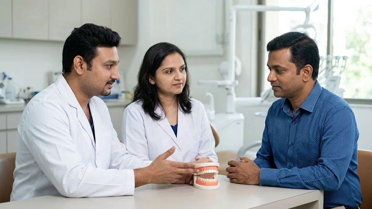
To prevent these devastating consequences, our dual-surgeon team—Dr. Pradeep S. and Dr. Kalpa Pandya—meticulously plans immediate reconstruction alongside the initial removal of the diseased tissue.
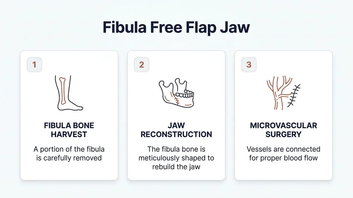
Fibula Free Flap: How Leg Bone Rebuilds Your Jaw and Holds Implants
The term "free flap" refers to a tissue transfer technique where bone, skin, muscle, and their supplying blood vessels are completely detached from one part of the body (the donor site) and transplanted to another (the recipient site). The blood vessels of the donor tissue are then microsurgically reconnected to blood vessels in the neck to restore immediate circulation.
Why the Fibula is the Perfect Donor Site
The fibula is the thin, outer bone of the lower leg running parallel to the tibia (shinbone). It serves as an ideal donor site for several key reasons:
- Length and Strength: The fibula provides up to 25 centimetres of straight, dense, cortical bone. This length allows surgeons to reconstruct even total or near-total jaw defects.
- Non-Weight-Bearing: The tibia carries approximately 90% of the body's weight. The fibula is largely non-weight-bearing, meaning a significant middle portion can be safely harvested without compromising your ability to stand, walk, or run.
- Excellent Blood Supply: The bone is nourished by the peroneal artery and its accompanying veins. This robust vascular network ensures the bone remains alive and healthy after transplantation.
- Two-Team Surgery: Because the leg is far from the head and neck, one surgical team (led by Dr. Pradeep S.) can harvest the leg bone while another team prepares the jaw area. This parallel workflow significantly reduces total anesthesia time.
The Role of Microvascular Surgery
For the transplanted leg bone to survive in its new home, it must have an active blood supply. This is achieved through microvascular surgery. Using an advanced surgical microscope and sutures thinner than a human hair, we connect the peroneal artery of the fibula graft to a branch of the external carotid artery in the neck, and the peroneal veins to the jugular vein system.
Once blood flows through these newly joined vessels, the transferred leg bone behaves exactly like living jawbone—it heals, remodels, and can successfully integrate with dental implants.
Step-by-Step: The Surgical Journey of a Fibula Free Flap
A successful oral reconstruction requires meticulous planning, advanced technology, and precise execution. Here is a step-by-step breakdown of how our team performs this complex procedure at Apollo Main Hospital, Greams Road, Chennai.
1. Virtual Surgical Planning (VSP)
Before entering the operating room, we use high-resolution CT scans of the patient’s head and lower legs to create 3D digital models. Using specialized software, we simulate the exact bone cuts required to remove the tumour and design custom cutting guides for both the leg and the jaw.
We also design a custom titanium reconstruction plate that is pre-bent to match the patient's original jaw anatomy. This Virtual Surgical Planning (VSP) ensures unmatched precision, shortens surgery times, and optimizes the final cosmetic and functional outcome.
2. Harvesting the Leg Bone
While the tumor removal is underway, the reconstructive team makes an incision on the outer side of the lower leg. We carefully expose the fibula bone, preserving at least 6 to 8 centimetres of bone at both the knee and ankle joints to maintain joint stability.
The required length of bone is isolated along with the peroneal artery and veins, plus a small strip of skin (a "skin paddle") if soft tissue is needed to replace missing lining inside the mouth or outer cheek.
3. Shaping the Jaw (Osteotomies)
The harvested straight fibula bone must be shaped to match the natural arch of the jaw. Using the 3D-printed cutting guides, we perform precise bone cuts (osteotomies) while keeping the underlying blood vessels intact. The bone segments are folded like an accordion to mimic the curve of the chin and jawline.
4. Securing the Bone and Connecting the Vessels
The shaped fibula bone is placed into the jaw defect and rigidly secured to the remaining natural jaw using the pre-bent titanium plates and screws.
Next, the microvascular phase begins. Under a high-powered operating microscope, we perform the vascular anastomosis—connecting the tiny blood vessels of the leg bone to the blood vessels in the neck. Once blood flow is established, we verify the health of the graft using specialized clinical monitoring techniques.
Restoring Your Smile: How the Reconstructed Jaw Holds Implants
Rebuilding the structure of the jaw is only the first half of the journey. True rehabilitation is complete only when the patient can chew, speak, and smile naturally. This requires restoring the missing teeth.
Because the fibula consists of thick, dense cortical bone, it provides an exceptionally stable environment for full-arch dental implants.
Primary vs. Secondary Implant Placement
Depending on the patient’s overall health, the nature of their disease, and the need for post-operative radiation therapy, dental implants can be placed in one of two ways:
- Primary (Immediate) Placement: In select cases, dental implants are inserted into the fibula bone during the very same surgery in which the jaw is rebuilt. This is often referred to as "Jaw-in-a-Day." It allows for faster overall dental rehabilitation but requires highly specialized, multi-disciplinary coordination.
- Secondary (Delayed) Placement: More commonly, especially if the patient requires post-operative radiation therapy for oral cancer, we allow the reconstructed jawbone to heal completely first. After approximately 6 to 12 months, once the bone has fully integrated and any oncology treatments are complete, Dr. Kalpa Pandya performs a minor secondary procedure to place the dental implants.
The Osseointegration Process in Leg Bone
Once dental implants (titanium posts that act as artificial tooth roots) are placed into the reconstructed fibula, a biological process called osseointegration occurs. Over a period of 3 to 4 months, the living leg bone fuses directly with the titanium surface of the implants.
Once osseointegration is complete, custom-made dental prostheses (crowns, bridges, or implant-supported dentures) are securely fixed to the implants, restoring near-normal biting force and beautiful dental aesthetics.
Comparing Jaw Reconstruction Options
While the fibula free flap is the most versatile option, other donor sites exist. The table below compares the primary reconstructive options used in modern maxillofacial surgery:
| Reconstructive Option | Primary Benefits | Best Suited For | Ability to Support Implants | Recovery Considerations |
|---|---|---|---|---|
| Fibula Free Flap | Long bone length, dual-team surgery, excellent bone density, consistent blood supply. | Large mandible or maxilla defects requiring multiple dental implants. | Excellent (Gold Standard) | Temporary leg weakness; requires physical therapy; no long-term mobility loss. |
| Iliac Crest (Hip) Free Flap | Natural curvature, abundant bone width and height. | Shorter jaw defects requiring significant height (e.g., matching a deep bite). | Good | Significant temporary post-operative hip pain and limp. |
| Scapula (Shoulder) Free Flap | Large amount of skin and soft tissue available; highly mobile bone. | Complex defects involving both jawbone and extensive facial skin/palate loss. | Moderate | Restricts shoulder mobility temporarily; requires intensive physical therapy. |
| PMMC Flap (Pectoralis Major) | Simple, fast, does not require microvascular surgery. | Patients unable to tolerate long microsurgical procedures; soft tissue reconstruction only. | No (Does not contain bone) | Limited to soft tissue bulk; cannot restore teeth. |
For more details on non-microvascular options, you can read about the PMMC flap procedure.
Recovery and Rehabilitation: What to Expect After Surgery
A fibula free flap is a major surgical undertaking. Understanding the recovery timeline helps patients and caregivers prepare mentally and physically for the journey ahead.
Phase 1: The Hospital Stay (Days 1 to 14)
- The ICU (First 24-48 Hours): Patients spend the first day or two in the intensive care unit. We closely monitor the blood flow to the newly reconstructed jaw every hour using specialized Doppler probes and clinical checks.
- Airway Management: Swallowing will be difficult initially. A temporary tracheostomy (a breathing tube in the neck) is often placed during surgery to protect your airway and is typically removed within 5 to 7 days.
- Nutrition: A temporary feeding tube (nasogastric tube) is used to provide liquid nutrition while the incisions inside the mouth heal.
- Leg Care: The leg donor site is kept in a splint or heavy dressing to protect the healing tissues.
Phase 2: Early Mobilization (Weeks 2 to 6)
- Walking Again: Physical therapy starts early—often within 3 to 4 days of surgery. Patients transition from dangling their legs to standing, and eventually walking with a walker or crutches. Most patients walk independently without assistance by week 6.
- Transitioning to Oral Diet: As swelling subsides, patients gradually transition from tube feeding to a pureed or soft diet.
- Speech Therapy: Gentle speech exercises help the tongue and lips adapt to the newly shaped jaw.
Phase 3: Long-Term Healing and Implants (Months 3 to 12)
- Bone Fusion: By month 3, the fibula bone has securely fused with the native jawbone.
- Dental Rehabilitation: If implants were not placed during the primary surgery, this is the window where we plan and execute implant placement, followed by the fabrication of custom teeth.
If you are currently experiencing symptoms or have been diagnosed with a condition requiring jaw surgery, early consultation is vital. Book an appointment with Dr. Pradeep S. and Dr. Kalpa Pandya at Apollo Main Hospital, Greams Road, Chennai, to discuss your reconstructive options.
Why Choose the Dual-Surgeon Team at Mouth Cancer Surgeons, Chennai
Reconstructive microsurgery of the face is highly complex and demands absolute precision. At Mouth Cancer Surgeons, we offer a unique, collaborative care model that ensures the highest standards of safety and success:
- The Power of Two: Dr. Pradeep S. and Dr. Kalpa Pandya work together on every major reconstructive case. While Dr. Pradeep S. focuses on the oncology clearance and the intricate microvascular reconstruction, Dr. Kalpa Pandya leads the precise planning of the jaw alignment, facial aesthetics, and dental implant rehabilitation.
- End-to-End Continuity: The same two surgeons who diagnose your condition will perform your surgery, manage your hospital recovery, and guide you through long-term dental rehabilitation and surveillance. You will never be passed off to rotating residents or junior doctors.
- World-Class Infrastructure: We perform our major reconstructive procedures at Apollo Main Hospital, Greams Road, Chennai—a premier healthcare facility equipped with state-of-the-art operating theatres, advanced surgical microscopes, and dedicated post-operative intensive care.
For personalized treatment options and expert care, consult Dr. Pradeep S. and Dr. Kalpa Pandya — Mouth Cancer Surgeons, Chennai. Call +91 96633 03747 or book an appointment online today.
References
- Sozzi, Davide, et al. "Virtual Surgical Planning and 3D Printing in Fibula Free Flap Jaw Reconstruction." Journal of Cranio-Maxillofacial Surgery, 2020.
- Wong, Christopher H., et al. "The Fibula Osteocutaneous Free Flap: Anatomy, Harvesting Techniques, and Clinical Applications." Plast Reconstr Surg, 2018.
- National Comprehensive Cancer Network (NCCN). "NCCN Clinical Practice Guidelines in Oncology: Head and Neck Cancers." 2024. NCCN Guidelines
- Shanmugam, S., et al. "Microvascular Free Flap Reconstruction in Oral Cancer: A South Indian Tertiary Care Experience." Indian Journal of Surgical Oncology, 2021.
- Association of Oral and Maxillofacial Surgeons of India (AOMSI). "Textbook of Oral and Maxillofacial Surgery." 2022.
Authored by
Medically reviewed by