Rebuilding the Jaw After Cancer: Free Flap vs PMMC Flap

Need expert consultation? Book an appointment with Dr. Pradeep S. or Dr. Kalpa Pandya.
Book AppointmentThe diagnosis of advanced oral cancer often brings with it the daunting prospect of major surgery. When a tumor invades the mandible (lower jaw) or the maxilla (upper jaw), surgical removal of the affected bone—a procedure known as a mandibulectomy or maxillectomy—becomes necessary to achieve clear oncological margins. While removing the cancer is the primary goal, restoring the structural integrity of the face, the ability to swallow, speak, and chew, is equally vital. For patients undergoing this procedure in Chennai, understanding the reconstructive options is a critical step in their treatment.
Rebuilding the jaw after cancer free flap vs PMMC flap represents the two primary surgical methodologies used by reconstructive surgeons. Each approach has distinct indications, advantages, and limitations. At our practice, Mouth Cancer Surgeons in Chennai, Dr. Pradeep S. and Dr. Kalpa Pandya utilize their combined expertise in head and neck oncology and maxillofacial reconstruction to tailor these procedures to each patient's unique anatomical and physiological needs.
Understanding Jaw Resection in Oral Cancer Surgery
Oral cancers, particularly squamous cell carcinomas of the lower alveolus (gums), buccal mucosa (inner cheek), or floor of the mouth, frequently invade the adjacent jawbone. When imaging studies, such as a contrast-enhanced CT scan or an orthopantomogram (OPG), reveal bone invasion, the surgical team must plan for bone resection.
Depending on the extent of the invasion, the surgeon may perform:
- Marginal Mandibulectomy: Removal of the upper border of the jawbone, preserving the continuity of the lower border of the mandible.
- Segmental Mandibulectomy: Complete removal of a segment of the jawbone, which breaks the continuity of the mandible.
A segmental resection completely disrupts the structural arch of the lower face. Without immediate reconstruction, this leads to severe cosmetic deformity, deviation of the remaining jaw, inability to chew, difficulty swallowing (dysphagia), and compromised airway protection.
To prevent these debilitating outcomes, reconstructive surgery is performed in the same surgical setting immediately following the tumor removal. This complex restorative phase is where the choice between a microvascular free flap and a regional pedicled flap, such as the Pectoralis Major Myocutaneous (PMMC) flap, becomes critical.
If you want to learn more about the broader classifications of oral malignancies, read our detailed guide on the types of oral cancer.
What is a Microvascular Free Flap Reconstruction?
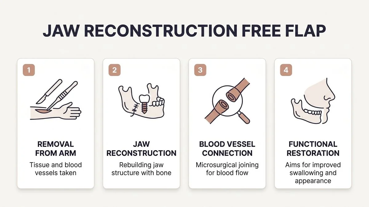
A microvascular free flap, often referred to simply as a "free flap," is the modern gold standard for complex jaw reconstruction. This advanced technique involves harvesting a tissue package (including bone, skin, muscle, or a combination of these) from a distant donor site on the patient's body, along with its primary feeding artery and draining veins.
This tissue is completely detached from its native blood supply, transferred to the facial defect, and its blood vessels are meticulously reconnected to recipient blood vessels in the neck using microvascular surgical techniques under an operating microscope.
For jaw reconstruction, the most common donor sites include:
- The Fibula Free Flap (FFF): Harvested from the calf bone, the fibula provides a long, straight segment of strong cortical bone that can be precisely cut (osteotomized) and shaped to mimic the natural curve of the mandible or maxilla. It also carries a skin paddle to reconstruct internal oral lining or external facial skin.
- The Deep Circumflex Iliac Artery (DCIA) Flap: Harvested from the hip bone, providing a natural curvature ideal for shorter, deeper jaw defects.
- The Radial Forearm Free Flap (RFFF) or Anterolateral Thigh (ALT) Flap: These are primarily soft-tissue flaps used when bone reconstruction is not required but extensive lining or bulk is needed.
To explore the details of how this complex procedure is planned and executed, visit our dedicated section on reconstructive & restorative surgery.
What is a Pectoralis Major Myocutaneous (PMMC) Flap?
The Pectoralis Major Myocutaneous (PMMC) flap is a regional, pedicled flap that has served as the "workhorse" of head and neck reconstruction since its introduction in the late 1970s. Unlike a free flap, the PMMC flap is not completely detached from the body.
Instead, a portion of the pectoralis major muscle along with overlying chest skin is harvested from the patient's chest. This tissue package remains attached to its vascular "pedicle"—the thoracoacromial artery and vein—which provides its blood supply. The flap is then tunneled under the skin of the chest and neck and rotated upward into the oral cavity or facial defect.
Because the PMMC flap consists of muscle and skin, it does not naturally contain bone. When used for jaw reconstruction, it is typically utilized in one of two ways:
- Soft Tissue Reconstruction Alone: Used to close the soft tissue defect in patients where bone reconstruction is either contraindicated or not feasible due to advanced age or poor medical fitness.
- Combined with a Titanium Reconstruction Plate: The titanium plate is used to bridge the bone gap and maintain the shape of the jaw, while the PMMC flap is wrapped around the plate to provide bulk, protect the hardware, and close the oral mucosal defect.
For a comprehensive overview of how this regional flap is utilized in clinical practice, refer to our detailed guide on the PMMC flap procedure.
Rebuilding the Jaw After Cancer: Free Flap vs PMMC Flap Compared
Choosing between a free flap and a PMMC flap involves weighing several clinical, anatomical, and patient-specific factors. The table below outlines the core differences between these two reconstructive modalities:
| Feature | Microvascular Free Flap (e.g., Fibula Flap) | PMMC Flap (with or without Reconstruction Plate) |
|---|---|---|
| Primary Composition | Vascularized bone, muscle, and skin. | Muscle and skin (no native bone). |
| Structural Alignment | Excellent; bone can be shaped to match natural jaw contours. | Poor without a metal plate; relies on a titanium plate for structure. |
| Dental Rehabilitation | Highly feasible; allows for dental implants after bone healing. | Very limited; implants cannot be placed in a metal plate or muscle tissue. |
| Surgical Complexity | High; requires microvascular anastomosis under a microscope. | Moderate; straightforward dissection and rotation without vascular suturing. |
| Operative Time | Typically longer (8 to 12 hours) due to microvascular work. | Shorter (4 to 6 hours), reducing anesthesia exposure. |
| Donor Site Morbidity | Leg weakness, gait changes (temporary), or forearm scarring. | Chest wall scar, breast asymmetry, restricted shoulder mobility. |
| Flap Failure Risk | 2% to 5% risk of total flap loss due to microvascular thrombosis. | Low risk of total loss; higher risk of partial skin necrosis or wound breakdown. |
| Long-Term Durability | Exceptional; living bone integrates and heals permanently. | Moderate; high risk of titanium plate exposure over time under radiation. |
Clinical Decision-Making: How Surgeons Choose the Right Flap
At our practice in Chennai, Dr. Pradeep S. and Dr. Kalpa Pandya evaluate each patient using a comprehensive, multidisciplinary approach. The decision of rebuilding the jaw after cancer free flap vs PMMC flap is never one-size-fits-all. It is guided by a careful analysis of several critical variables.
1. The Nature and Location of the Defect
If the surgical resection results in a large segmental defect of the anterior mandible (the "chin" area), a free fibula flap is almost always preferred. Reconstructing the anterior jaw with a titanium plate and a PMMC flap alone often leads to a high rate of plate exposure, extrusion, and the classic "plate-through-skin" complication due to the constant mechanical stress of swallowing and speaking.
Conversely, for posterior defects (near the angle of the jaw or the ascending ramus) in elderly patients, a PMMC flap wrapping a reconstruction plate may provide an acceptable functional and cosmetic outcome with lower surgical risk.
2. Patient Age and Systemic Comorbidities
Microvascular free flap surgery is a lengthy, physically demanding procedure that requires stable cardiovascular and respiratory function to tolerate prolonged anesthesia. For patients with severe heart disease, uncontrolled diabetes, advanced peripheral vascular disease (which affects the blood vessels in the legs), or significant renal impairment, a shorter, less invasive PMMC flap procedure is often the safer choice.
3. Prior Radiation Therapy and Vessel Availability
Free flaps require healthy, patent recipient blood vessels in the neck (such as the facial artery and external jugular vein) for successful microvascular anastomosis. If a patient has undergone previous neck dissections or high-dose radiation therapy, these vessels may be severely scarred or thrombosed. In such cases, the PMMC flap, which carries its own blood supply from the chest, serves as an invaluable and highly reliable alternative.
4. Patient Goals and Long-Term Quality of Life
For younger, active patients, the primary goal is often complete functional restoration, including the ability to chew solid food. This requires a vascularized bone graft that can support dental implants. For these patients, the extra operative time and recovery associated with a fibula free flap are well justified.
For elderly patients whose primary goal is wound healing, pain control, and early return to a soft diet, a PMMC flap offers a faster pathway to recovery.
If you are concerned about the financial aspects of these complex procedures, you can read our detailed breakdown of how much oral cancer treatment costs in Chennai.
Postoperative Care and Flap Monitoring
The success of any reconstructive surgery depends heavily on the quality of postoperative care. At Apollo Main Hospital, Greams Road, Chennai, our patients are monitored in a specialized surgical intensive care unit (ICU) by a dedicated team of nurses and maxillofacial surgeons.
Monitoring a Free Flap
The first 72 hours after a microvascular free flap are the most critical. The tiny blood vessels sutured during surgery are at risk of developing blood clots (thrombosis). If a clot blocks the artery, the flap loses its oxygen supply; if it blocks the vein, the flap becomes severely congested.
To detect these issues early, the surgical team performs hourly clinical checks, assessing:
- Color: A healthy flap is pink; a pale flap indicates arterial insufficiency, while a bluish or purple flap indicates venous congestion.
- Temperature: Free flaps are monitored using surface temperature probes. A sudden drop in temperature suggests a vascular compromise.
- Capillary Refill: Pressing on the skin paddle of the flap should produce a rapid return of color (within 1 to 2 seconds).
- Handheld Doppler Ultrasound: Used to listen to the characteristic acoustic signals of arterial and venous blood flow within the flap.
If vascular compromise is detected, the patient is immediately returned to the operating theatre for microvascular re-exploration to clear the clot and rescue the flap.
Monitoring a PMMC Flap
Because the PMMC flap remains attached to its native blood supply, it does not require microscopic vascular monitoring. However, the surgical team must still monitor the surgical sites for:
- Venous Congestion: Occurs if the pedicle is twisted or compressed under the neck skin.
- Distal Necrosis: The edges of the chest skin paddle furthest from the blood supply may occasionally experience poor perfusion, leading to minor wound breakdown.
- Seroma or Hematoma: Fluid accumulation in the chest donor site, which is managed using active suction drains.
Restoring Function: Dental Implants and Speech Therapy
The ultimate goal of rebuilding the jaw is not merely to close the wound, but to restore the patient's quality of life. This phase of rehabilitation highlights the significant advantage of the fibula free flap.
Dental Rehabilitation
Because the fibula free flap provides living, vascularized bone, it undergoes normal bone healing and integrates with the remaining native jawbone. After a healing period of approximately 6 months, dental implants can be surgically placed directly into the reconstructed bone.
These implants then support custom-made dental prostheses (bridges or dentures), allowing the patient to chew a normal diet.
In contrast, a PMMC flap combined with a titanium reconstruction plate does not provide a bony foundation. Dental implants cannot be placed into muscle tissue or a metal plate. Patients reconstructed with this method must rely on a modified soft diet long-term.
To learn more about the possibilities of tooth replacement after reconstructive surgery, explore our specialized services in dental implants & pre-prosthetic rehabilitation.
[Infographic Placeholder]
rebuilding the jaw after cancer free flap vs PMMC flap — key facts infographic
Speech and Swallowing Therapy
Regardless of the flap chosen, extensive tissue rearrangement in the oral cavity affects speech articulation and swallowing mechanics. Early intervention by a speech and language pathologist is essential.
Exercises designed to improve tongue mobility, lip closure, and laryngeal elevation help patients regain clear speech and transition safely from tube feeding to oral intake.
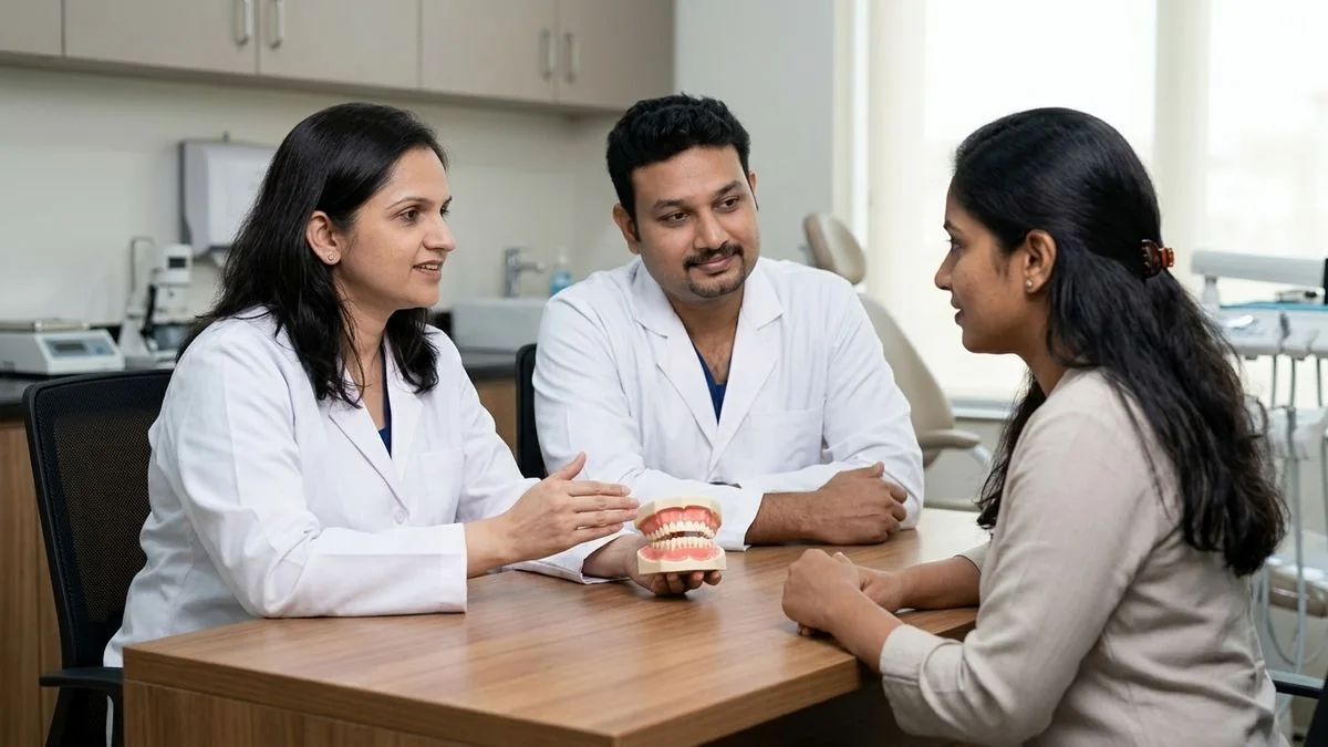
The Dual-Surgeon Advantage for Complex Reconstruction
At Mouth Cancer Surgeons, Chennai, we operate under a unique, highly collaborative dual-surgeon model. Dr. Pradeep S. and Dr. Kalpa Pandya work together on every complex reconstructive case, providing a seamless continuum of care from diagnosis to final rehabilitation.
- Dr. Pradeep S. brings extensive super-specialty expertise in head and neck surgical oncology and microvascular reconstruction. He leads the tumor ablation phase, ensuring clean surgical margins, and directs the complex microvascular harvesting and anastomosis.
- Dr. Kalpa Pandya specializes in oral and maxillofacial surgery, OPMD management, and advanced dental implant rehabilitation. She leads the precise shaping of the reconstructed bone, ensures optimal jaw alignment (occlusion), and directs the long-term dental implant phase.
By having both surgeons present in the operating room, we drastically reduce operative times—a crucial factor in minimizing anesthesia-related complications for elderly or medically compromised patients. While one surgeon focuses on removing the tumor and preparing the neck vessels, the other simultaneously harvests the reconstructive flap, ensuring maximum efficiency and safety.
Our primary surgical consultations and procedures are conducted at Apollo Main Hospital, Greams Road, Chennai, a tertiary care center equipped with state-of-the-art surgical ICUs, advanced operating microscopes, and comprehensive multidisciplinary oncology support.
If you or a loved one are facing oral cancer surgery and wish to discuss your reconstructive options, early planning is key to achieving the best functional outcomes.
For personalized treatment options and expert care, consult Dr. Pradeep S. and Dr. Kalpa Pandya — Mouth Cancer Surgeons, Chennai. Call +91 96633 03747 or book an appointment.
References
- Urken, M. L., et al. "Primary reconstruction of the mandible with microvascular free flaps: Analysis of 10 cases." Laryngoscope, 1989.
- Ariyan, S. "The pectoralis major myocutaneous flap. A versatile flap for reconstruction in the head and neck." Plastic and Reconstructive Surgery, 1979.
- Hidalgo, D. A. "Fibula free flap: A new method of mandible reconstruction." Plastic and Reconstructive Surgery, 1989.
- National Comprehensive Cancer Network (NCCN). "Clinical Practice Guidelines in Oncology: Head and Neck Cancers." 2025.
- Kroll, S. S., et al. "A comparison of resource utilization and clinical outcomes of free flaps and pedicled flaps in head and neck reconstruction." Plastic and Reconstructive Surgery, 1997.
Authored by
Medically reviewed by