Oral Cancer Stages Explained: Early to Advanced Chennai
Need expert consultation? Book an appointment with Dr. Pradeep S. or Dr. Kalpa Pandya.
Book AppointmentReceiving a diagnosis or experiencing symptoms that point toward oral cavity cancer can be overwhelming. Understanding how the disease is staged is one of the most critical steps in reclaiming control over your health. Staging is not merely a label; it is a highly structured, scientific roadmap that oral and maxillofacial oncologists use to design your surgical plan, determine the necessity of reconstructive procedures, and estimate overall prognosis.
When we explain the progression of oral cancer to our patients at our Chennai practice, we emphasize that early detection completely transforms the treatment landscape. This detailed guide breaks down oral cancer stages explained from early lesion to advanced, detailing how we evaluate, classify, and treat each phase of the disease.
What is Oral Cancer Staging and Why Does It Matter?
Oral cancer staging is the process of finding out how much cancer is in the body and where it is located. We primary stage cancers of the oral cavity, which includes the tongue, buccal mucosa (inner cheek), floor of the mouth, alveolus (gums), hard palate, and the lips.
The gold standard for staging is the American Joint Committee on Cancer (AJCC) TNM system. This system evaluates three key parameters:
- T (Tumor): The size of the primary tumor and how deeply it has grown into local tissues (Depth of Invasion).
- N (Node): Whether the cancer has spread to the lymph nodes in the neck, including the number, size, and location of these nodes.
- M (Metastasis): Whether the cancer has spread to distant parts of the body, such as the lungs, liver, or bones.
The Critical Role of Depth of Invasion (DOI)
In the updated AJCC 8th edition, the depth of invasion (DOI) was introduced as a major staging factor for oral cavity cancers. DOI measures how deeply the tumor has penetrated beneath the mucosal surface. A small tumor on the surface (e.g., less than 2 cm) that penetrates deeply (more than 5 mm) into the underlying muscle is classified as a more aggressive stage than a flat surface lesion of the same width. This is because a deeper invasion significantly increases the likelihood that cancer cells have traveled to the lymph nodes in the neck.

The Precursor Phase: Oral Potentially Malignant Disorders (OPMD)
Before a true cancer develops, abnormal tissue changes often occur in the mouth. These are classified as Oral Potentially Malignant Disorders (OPMD). While not cancer yet, these lesions have a statistical risk of transforming into squamous cell carcinoma (the most common type of oral cancer).
Common OPMDs include:
- Leukoplakia: White patches in the mouth that cannot be rubbed off or characterized as any other disease.
- Erythroplakia: Red, velvety patches that carry a much higher risk of malignancy than leukoplakia.
- Oral Submucous Fibrosis (OSMF): A chronic, progressive condition characterized by mucosal rigidity and a progressive inability to open the mouth, highly associated with betel nut chewing.
In our practice, we closely monitor and biopsy these lesions. Catching an OPMD and treating it through conservative surgical excision or lifestyle modification can prevent the development of invasive oral cancer entirely. If you notice persistent red or white spots, reviewing them early is vital. Read more about identifying these early signs in our article on mouth ulcer warning signs.
Stage 0: Carcinoma in Situ (Pre-Invasive Cancer)
Stage 0 is also known as carcinoma in situ (CIS). At this stage, highly atypical, cancerous cells are present, but they are entirely confined to the superficial epithelial layer of the oral mucosa. They have not broken through the basement membrane to invade the deeper layers of tissue, blood vessels, or lymphatic channels.
- Clinical Presentation: Often presents as a non-healing red or white patch that may feel slightly rough or sensitive but is rarely painful.
- Treatment: Stage 0 is highly curable. The primary treatment is a wide local excision with clear margins. Because the disease is superficial, major reconstruction is rarely required, and the regional lymph nodes do not need to be surgically removed.
Early-Stage Oral Cancer: Stages I and II
Stages I and II represent localized oral cancers. The cancer has invaded past the basement membrane into the deeper tissues of the mouth, but it remains confined to the local site of origin without spreading to the lymph nodes.
Stage I Oral Cancer
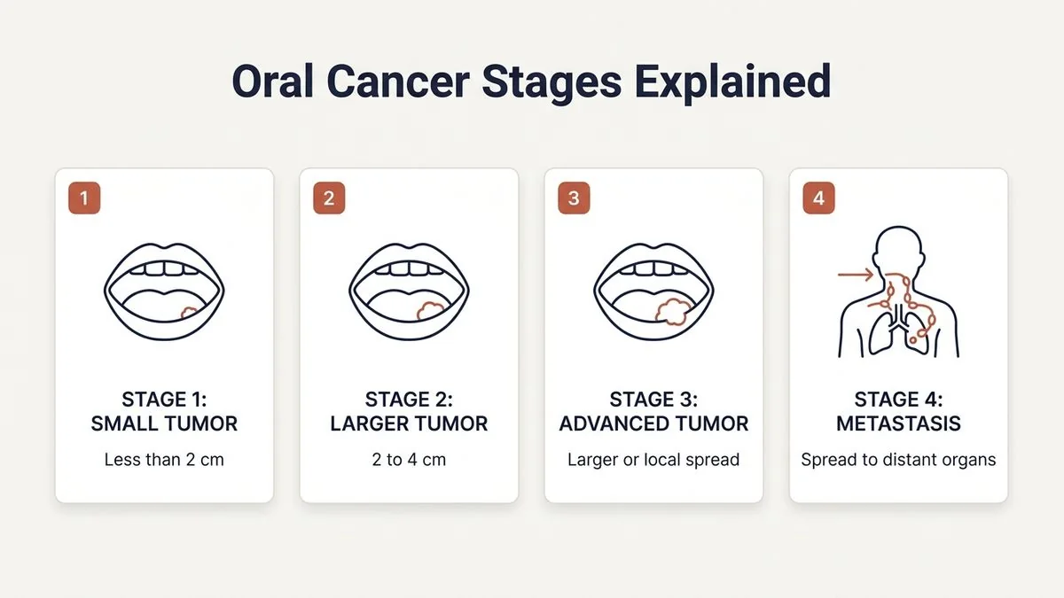
In Stage I, the tumor is relatively small and superficial:
- The tumor size is 2 cm or smaller.
- The Depth of Invasion (DOI) is 5 mm or less.
- There is no spread to regional lymph nodes (N0) or distant sites (M0).
Stage II Oral Cancer
In Stage II, the cancer has grown larger or penetrated deeper:
- The tumor size is 2 cm or smaller with a DOI of more than 5 mm and up to 10 mm; OR
- The tumor is between 2 cm and 4 cm in size with a DOI of 10 mm or less.
- There is no spread to regional lymph nodes (N0) or distant sites (M0).
Treatment for Early-Stage Oral Cancer
The primary treatment for Stage I and Stage II oral cancer is surgical resection. Dr. Pradeep S. and Dr. Kalpa Pandya perform precise oral cancer surgery to excise the tumor with a surrounding margin of healthy tissue to ensure no microscopic cancer cells are left behind.
Even in early stages, if the DOI is greater than 3 to 4 mm, we often recommend an elective neck dissection (removal of lymph nodes in the neck). This is a preventive measure because subclinical, microscopic cancer spread to the neck nodes can occur even when scans show no visible swelling.
Clinical Insight: Early-stage oral cancers have an excellent prognosis, with 5-year survival rates often exceeding 85%. Seeking a specialist consultation immediately upon noticing a non-healing ulcer or growth is the single most effective action a patient can take.
Locally Advanced Oral Cancer: Stage III
Stage III oral cancer represents a transition from a localized disease to a locally advanced disease. It is defined by either a larger primary tumor or the initial spread of cancer cells to a single lymph node on the same side of the neck.
A cancer is classified as Stage III if it meets any of the following criteria:
- The tumor is larger than 4 cm in its greatest dimension; OR
- The tumor has a Depth of Invasion (DOI) greater than 10 mm, regardless of its surface diameter; OR
- The tumor is of any size but has spread to a single lymph node on the same side of the neck, and that lymph node is 3 cm or smaller in size.
Clinical Implications of Stage III
At this stage, patients often experience more pronounced symptoms, such as persistent pain, difficulty swallowing (dysphagia), changes in speech, or a palpable, painless lump in the neck.
Treatment for Stage III oral cancer is multidisciplinary. It almost always involves:
- Radical Surgical Resection: Removing the primary tumor with wide margins.
- Therapeutic Neck Dissection: Surgical removal of the lymph nodes in the neck to clear any nodal metastasis.
- Reconstructive Surgery: Using advanced tissue transfer techniques to restore speech, swallowing, and facial appearance.
- Adjuvant Therapy: Radiation therapy, often combined with chemotherapy, to eliminate any remaining microscopic cancer cells and reduce the risk of recurrence.
If you are experiencing progressive swelling, pain, or difficulty swallowing, early expert evaluation is critical. Book an appointment with Dr. Pradeep S. and Dr. Kalpa Pandya at Apollo Main Hospital, Greams Road, Chennai.
Advanced Oral Cancer: Stage IV
Stage IV is the most advanced stage of oral cancer. It is sub-classified into Stage IVA, IVB, and IVC based on the extent of local tissue invasion, the severity of lymph node involvement, and whether distant organs are affected.
[Stage IV Sub-Classifications]
  ├── Stage IVA (Moderately Advanced Local / Regional Disease)
  │     └── Invades adjacent structures (jawbone, skin) OR multiple/large neck nodes.
  ├── Stage IVB (Very Advanced Local / Regional Disease)
  │     └── Invades deep skull base, carotid artery, or extensive fixed neck nodes.
  └── Stage IVC (Distant Metastatic Disease)
        └── Cancer has spread to distant organs (lungs, liver, bones).
Stage IVA: Moderately Advanced Local or Regional Disease
This stage indicates that the cancer has either invaded adjacent structures or spread extensively to the neck lymph nodes, but has not traveled to distant organs.
- Local Invasion: The tumor has grown through the oral cavity to invade adjacent structures such as the cortical bone of the mandible (jawbone) or maxilla, the skin of the face, the deep extrinsic muscles of the tongue, or the maxillary sinus.
- Regional Lymph Nodes: Alternatively, the tumor may be smaller, but it has spread to multiple lymph nodes on the same side of the neck, to lymph nodes on both sides of the neck, or to a single lymph node larger than 3 cm but not exceeding 6 cm.
Stage IVB: Very Advanced Local or Regional Disease
This represents a highly advanced stage where surgical removal of the tumor becomes technically challenging or impossible due to the involvement of critical anatomical structures.
- The tumor invades the masticator space, pterygoid plates, or skull base; OR
- The tumor encases the internal carotid artery; OR
- The neck lymph node disease is extensive, featuring nodes larger than 6 cm or nodes that have become fixed to the surrounding deep neck tissues.
Stage IVC: Distant Metastatic Disease
Stage IVC is diagnosed when the oral cancer has spread through the bloodstream to distant organs. The most common site of distant metastasis for head and neck cancers is the lungs, followed by the liver and bones. At this stage, the primary goal of treatment shifts from curative surgery to systemic therapies (chemotherapy, immunotherapy) and palliative care to manage symptoms and prolong quality of life.
How Surgeons Determine the Stage: The Diagnostic Workup
To accurately stage oral cancer before initiating treatment, our dual-surgeon team at Mouth Cancer Surgeons, Chennai, performs a meticulous diagnostic workup. Accurate staging prevents under-treating aggressive cancers and avoids over-treating early-stage, localized lesions.
The staging process includes:
- Clinical Examination: A thorough visual and tactile examination of the entire oral cavity, pharynx, and neck. We feel the neck tissues to check for enlarged or firm lymph nodes.
- Tissue Biopsy: The definitive diagnosis. A small sample of the suspicious tissue is removed and examined under a microscope by a specialized head and neck pathologist to confirm the presence of squamous cell carcinoma.
- High-Resolution Imaging:
- Contrast-Enhanced CT (CECT): Excellent for evaluating bone invasion in the jaw (mandible or maxilla) and identifying enlarged neck lymph nodes.
- Magnetic Resonance Imaging (MRI): The gold standard for assessing soft tissue tumors, particularly tongue cancers and buccal mucosa cancers, as it clearly delineates the Depth of Invasion (DOI).
- PET-CT Scan: Indicated for advanced stages (Stage III and IV) to screen the entire body for distant metastases and evaluate regional nodal disease.
- Endoscopy: An endoscopic evaluation of the upper aerodigestive tract may be performed to ensure there are no synchronous primary tumors in the throat or esophagus.
Survival Rates and Prognosis Across Stages
Understanding survival statistics can be difficult, but they provide essential context regarding the importance of early intervention. The survival of oral cancer is highly dependent on the stage at diagnosis.
The following table outlines the approximate 5-year relative survival rates for oral cavity cancers based on data from the AJCC and the SEER (Surveillance, Epidemiology, and End Results) database:
| Stage Grouping | SEER Stage Equivalent | Description | 5-Year Relative Survival Rate |
|---|---|---|---|
| Stage I & II | Localized | Cancer is confined entirely to the primary site in the oral cavity. | 83% – 90% |
| Stage III & IVA/B | Regional | Cancer has spread to regional lymph nodes or invaded adjacent tissues. | 60% – 65% |
| Stage IVC | Distant | Cancer has metastasized to distant organs (e.g., lungs). | 38% – 40% |
Note: These statistics are general aggregates. Individual prognosis depends heavily on tumor biology, the patient's general health, and the expertise of the surgical and oncological team. To understand how personalized factors influence outcomes, read our comprehensive article on is mouth cancer curable.
Stage-Based Treatment Pathways
At Mouth Cancer Surgeons, Chennai, Dr. Pradeep S. and Dr. Kalpa Pandya design customized treatment plans tailored to the precise stage of your disease. We operate as a cohesive dual-surgeon team, meaning the same two specialists plan, perform, and follow up on your surgery.
Treatment for Early Stages (Stage I & II)
- Surgery: Wide local excision of the primary tumor.
- Neck Management: Elective neck dissection is performed if the tumor thickness/DOI exceeds 3-4 mm.
- Reconstruction: Minimal local tissue rearrangement or primary closure is typically sufficient.
- Adjuvant Therapy: Rarely required unless pathological examination reveals close margins or unexpected aggressive features.
Treatment for Advanced Stages (Stage III & IVA)
- Surgery: Radical resection of the primary tumor, which may involve removing portions of the jawbone (segmental or marginal mandibulectomy) or a portion of the tongue (hemiglossectomy or subtotal glossectomy).
- Neck Management: Comprehensive neck dissection to remove all at-risk lymph nodes.
- Reconstruction: Immediate reconstructive and restorative surgery using microvascular free flaps (e.g., fibula free flap for jaw reconstruction, radial forearm free flap for tongue/cheek reconstruction). This is essential to preserve facial symmetry, speech, and swallowing function.
- Adjuvant Therapy: Post-operative radiation therapy, often combined with chemotherapy (chemoradiotherapy), is initiated within 4 to 6 weeks of surgery to target any microscopic residual disease.
Reconstructive and Restorative Care: Life After Cancer Surgery
For patients diagnosed with Stage III or Stage IV oral cancer, the fear of facial disfigurement and loss of function is often as significant as the fear of the cancer itself. Modern reconstructive surgery has revolutionized the recovery process.
Our dual-surgeon practice specializes in immediate microvascular reconstruction. When Dr. Pradeep S. leads the oncological resection to remove the cancer completely, Dr. Kalpa Pandya simultaneously prepares the reconstructive phase, ensuring minimal operating time and optimal aesthetic and functional outcomes.
- Jaw Reconstruction: Using a vascularized segment of the fibula (leg bone) to rebuild a removed jawbone.
- Soft Tissue Reconstruction: Rebuilding the tongue or inner cheek using skin and muscle flaps from the arm, thigh, or chest.
- Dental Rehabilitation: Once the reconstructed bone has healed, we can place dental implants directly into the reconstructed jaw, restoring the patient’s ability to chew, speak, and smile naturally.
For a detailed breakdown of the financial aspects of these advanced procedures, you can read our guide on oral cancer treatment cost.
Seeking Expert Care in Chennai
If you or a loved one has been diagnosed with oral cancer, or if you are experiencing persistent symptoms like a non-healing ulcer, red or white patches, or difficulty opening your mouth, seeking an immediate expert consultation is paramount.
At Mouth Cancer Surgeons, Chennai, we offer a dedicated, compassionate, and highly specialized dual-surgeon model. Dr. Pradeep S. and Dr. Kalpa Pandya combine their extensive expertise in head and neck surgical oncology and microvascular reconstruction to provide world-class care at Apollo Main Hospital, Greams Road, Chennai.
By choosing our practice, you benefit from:
- Dual-Surgeon Expertise: Two senior oral and maxillofacial surgeons reviewing and executing your treatment plan together.
- Comprehensive Care: Seamless management from initial staging biopsy, through complex resection, microvascular reconstruction, and long-term surveillance.
- State-of-the-Art Infrastructure: Access to the advanced diagnostic, surgical, and intensive care facilities at Apollo Main Hospital, Greams Road.
For personalized treatment options and expert care, consult Dr. Pradeep S. and Dr. Kalpa Pandya — Mouth Cancer Surgeons, Chennai. Call +91 96633 03747 or book an appointment.
References
- Amin, Mahul B., et al. "The Eighth Edition AJCC Cancer Staging Manual: Continuing to build a bridge from a population-based to a more 'personalized' approach to cancer staging." CA: A Cancer Journal for Clinicians, 2017.
- Lydiatt, Daniel D., et al. "Head and Neck cancers—major changes in the American Joint Committee on Cancer eighth edition cancer staging manual." CA: A Cancer Journal for Clinicians, 2017.
- National Comprehensive Cancer Network (NCCN). "NCCN Clinical Practice Guidelines in Oncology: Head and Neck Cancers." NCCN, 2024. [https://www.nccn.org]
- World Health Organization. "Classification of Tumours: Head and Neck Tumours." IARC Press, 2024.
- National Cancer Institute. "SEER Cancer Statistics Review." NCI, 2023. [https://seer.cancer.gov]
Authored by

Medically reviewed by