White Spots on Lips: Benign Causes & When to Worry Chennai

Need expert consultation? Book an appointment with Dr. Pradeep S. or Dr. Kalpa Pandya.
Book AppointmentDiscovering an unexpected change in your mouth can be highly distressing. When patients visit our practice at Apollo Main Hospital on Greams Road, Chennai, one of the most common concerns they present with is the sudden appearance of pale or white spots on lips benign causes Fordyce milia when to worry are often top of mind during these consultations. While many of these lesions are entirely harmless, some can serve as early warning signs of more significant mucosal conditions.
Understanding the differences between benign anatomical variants and potentially serious pathological changes is essential. This clinical guide provides a comprehensive overview of why white spots form on the lips, how to identify harmless conditions like Fordyce spots and milia, and when it is critical to seek an expert evaluation from an oral and maxillofacial surgeon.
Understanding Lip Anatomy and Why White Spots Form
The lips are highly specialized structures that mark the transition from the external facial skin to the moist internal oral mucosa. This transitional zone is known as the vermilion border. Because of this unique histological makeup, the lips are susceptible to dermatological conditions, glandular anomalies, and mucosal diseases.
The outer lip is covered by keratinized stratified squamous epithelium, which contains hair follicles, sebaceous (oil) glands, and sweat glands. The inner lip is lined by non-keratinized labial mucosa, rich in minor salivary glands that keep the mouth lubricated. The vermilion border itself is thin, translucent, and lacks sweat glands or hair follicles, making underlying blood vessels highly visible (giving the lips their red color).
When white spots appear on this delicate tissue, they generally stem from one of four pathological or anatomical mechanisms:
- Glandular Anomalies: Ectopic sebaceous glands that become visible through the thin epithelium.
- Keratin Retention: Trapping of dead skin proteins (keratin) beneath the mucosal or cutaneous surface.
- Infections: Localized viral, bacterial, or fungal proliferation affecting the epithelial cells.
- Epithelial Changes (Dysplasia/Hyperkeratosis): Abnormal thickening of the keratin layer or cellular changes within the epithelium, which can block the red color of the underlying blood vessels, presenting as a permanent white patch.
Common Benign Causes of White Spots on Lips
The vast majority of white spots on the lips are benign, non-contagious, and do not require aggressive surgical intervention. In our clinical practice, we categorize these benign lesions to help reassure patients while maintaining a high level of diagnostic vigilance.
Fordyce Spots (Sebaceous Prominences)
Fordyce spots are the single most common cause of tiny white or yellowish-white bumps on the lips. Histologically, these are ectopic sebaceous glands. While sebaceous glands are normally associated with hair follicles on the skin, Fordyce spots are "ectopic" because they occur on hairless mucosal surfaces.
- Appearance: They present as tiny, discrete, painless papules measuring 1 to 3 millimeters in diameter. They often appear in large clusters or symmetrical bands along the vermilion border of the upper lip or on the inside of the cheeks (buccal mucosa).
- Sensation: They are completely asymptomatic. They do not itch, hurt, or bleed.
- Clinical Significance: Fordyce spots are a normal anatomical variant, present in up to 80% of adults. They are not infectious, are not sexually transmitted, and do not carry any risk of malignant transformation. Treatment is rarely necessary and is only sought for cosmetic reasons using laser ablation or micro-punch excision.
Milia
Milia are small, firm, dome-shaped white cysts that develop on the skin surface, including the cutaneous borders of the lips.
- Appearance: They look like tiny, pearly-white or yellowish beads. Unlike Fordyce spots, which are deeply integrated into the mucosal tissue, milia feel like tiny, hard seeds sitting just beneath the surface.
- Mechanism: Milia occur when dead skin cells and keratin become trapped in a miniature pocket or hair follicle infundibulum rather than shedding naturally.
- Clinical Significance: They are entirely benign. While they are highly common in newborns (often called "milk spots"), they can occur at any age. They frequently resolve on their own, though persistent lesions can be safely expressed or treated with minor dermatological procedures.
Oral Thrush (Pseudomembranous Candidiasis)
Oral thrush is a fungal infection caused by an overgrowth of Candida albicans, a yeast that naturally resides in the oral cavity in small amounts.
- Appearance: It presents as creamy white patches on the inner lips, tongue, palate, and buccal mucosa. These patches have a unique "cottage cheese" texture.
- Diagnostic Key: Unlike most other white lesions, the white plaques of oral thrush can be gently wiped or scraped off with a tongue depressor, leaving behind a raw, red, and sometimes bleeding mucosal surface.
- Sensation: Patients often complain of a cottony feeling in the mouth, loss of taste, or a mild burning sensation, especially when eating spicy or acidic foods.
- Risk Factors: It is commonly seen in individuals with weakened immune systems, those using steroid inhalers for asthma, diabetics, denture wearers, or patients who have recently completed a course of broad-spectrum antibiotics.
Herpes Simplex Virus (Cold Sores)
The Herpes Simplex Virus Type 1 (HSV-1) is a highly contagious viral infection that primarily affects the lips and surrounding tissues.
- Appearance: While fully formed cold sores are vesicular (fluid-filled blisters) that crust over, they often begin as small, pale, tight white bumps or micro-vesicles on the vermilion border.
- Sensation: The appearance of these spots is preceded by a highly characteristic prodromal phase consisting of tingling, itching, or a burning sensation on the specific spot of the lip.
- Clinical Significance: Once the vesicles rupture, they form painful, raw ulcers that crust over and heal within 7 to 14 days. Because it is a viral infection, active lesions contain live virus particles and are highly contagious.
Canker Sores (Aphthous Ulcers)
Aphthous ulcers are non-contagious inflammatory lesions that can develop on the inner, non-keratinized surfaces of the lips.
- Appearance: They present as round or oval sores with a distinct white, grey, or yellowish necrotic center surrounded by a highly defined, inflamed red border.
- Sensation: They are intensely painful, particularly during eating, speaking, or drinking.
- Clinical Significance: Most minor canker sores heal spontaneously within 10 to 14 days without scarring. They are often triggered by minor local trauma (such as an accidental bite), stress, nutritional deficiencies (Vitamin B12, iron, folic acid), or certain food sensitivities.
Distinguishing Benign Causes from Serious Conditions
To help you understand how benign anomalies compare to more significant mucosal conditions, our surgical team has compiled this comparative reference:
| Condition | Primary Appearance | Pain / Sensation | Scrapable? | Malignancy Risk | Recommended Action |
|---|---|---|---|---|---|
| Fordyce Spots | Tiny (1-3mm) yellowish-white clusters along the vermilion border. | None (completely asymptomatic). | No | 0% (Entirely benign anatomical variant) | Reassurance; no medical treatment required. |
| Milia | Hard, pearly-white dome-shaped tiny cysts on the outer lip border. | None. | No | 0% (Benign keratin retention) | Observation; cosmetic extraction if desired. |
| Oral Thrush | Creamy white, curd-like patches on inner lips and oral mucosa. | Mild burning, altered taste, dry mouth. | Yes (leaves a raw, red surface) | Very Low (unless systemic immune compromise) | Consult a specialist for topical antifungal therapy. |
| Cold Sores (HSV-1) | Tiny clusters of fluid-filled vesicles that turn into crusty sores. | Tingling, itching, burning before outbreak. | No (ruptures into fluid) | 0% (Viral infection) | Topical or oral antiviral medications; avoid contact. |
| Leukoplakia | Flat or slightly raised, thick, rough white patch on the lip. | Usually painless; may feel rough or firm. | No | Moderate to High (Potentially malignant) | Urgent biopsy and evaluation by an oral surgeon. |
| Actinic Cheilitis | Dry, scaly, white-to-greyish patches on the lower lip. | Dryness, scaling, occasional cracking. | No | High (Precursor to Squamous Cell Carcinoma) | Strict sun protection, biopsy of persistent thick areas. |
When to Worry: Red Flags and Warning Signs of Oral Precancer
While benign white spots are common, persistent white lesions on the lips can sometimes be classified as Oral Potentially Malignant Disorders (OPMD). Identifying these early is critical to preventing progression to invasive oral cancer.
If you notice a white spot on your lip, you should seek immediate evaluation from an oral and maxillofacial surgeon if you observe any of the following "red flag" characteristics:
- Persistence Beyond Two Weeks: Any white spot, patch, or ulcer on the lip that does not resolve or show significant healing within 14 days requires professional evaluation.
- Induration (Firmness): If the white area feels hard, firm, or thick when rolled between your fingers, rather than soft and pliable like the rest of your lip tissue.
- Inability to Scrape Off: If the white patch is fixed to the tissue and cannot be wiped or scraped away (unlike oral thrush).
- Associated Ulceration or Bleeding: If the white spot develops a central sore, cracks open, or bleeds spontaneously or with minor contact.
- Rapid Growth or Changing Margins: If the lesion is growing quickly, changing shape, or developing irregular, fuzzy, or speckled red-and-white borders (erythroleukoplakia).
- Associated Lymph Node Swelling: If you feel firm, painless, or swollen lumps in your neck or under your jaw alongside the lip spot.
Key Precancerous Conditions to Watch For
1. Leukoplakia of the Lip
Leukoplakia is a clinical term used to describe a white patch or plaque on the oral mucosa or lips that cannot be characterized clinically or pathologically as any other disease.
It is strongly associated with chronic irritation, tobacco use (both smoking and smokeless tobacco), and alcohol consumption. Histologically, leukoplakia can exhibit varying degrees of epithelial dysplasia (abnormal cellular changes). If dysplasia is present, the risk of the lesion transforming into oral squamous cell carcinoma is significantly elevated.
2. Actinic Cheilitis
Often referred to as "sailor's lip" or "farmer's lip," actinic cheilitis is a pre-malignant condition caused by long-term, chronic exposure to solar ultraviolet (UV) radiation. It almost exclusively affects the lower lip due to its anatomical position, which receives more direct sunlight than the upper lip.
- Presentation: It begins as a loss of the sharp border between the red part of the lip and the facial skin. The lip becomes chronically dry, scaly, and develops thin, white-to-greyish plaques. Over time, these plaques can thicken, crack, and form persistent ulcers.
- Malignant Potential: If left untreated, actinic cheilitis can progress directly into invasive squamous cell carcinoma of the lip.
If you are experiencing any of these persistent symptoms, early consultation is important. Book an appointment with Dr. Pradeep S. and Dr. Kalpa Pandya at Apollo Main Hospital, Greams Road, Chennai.
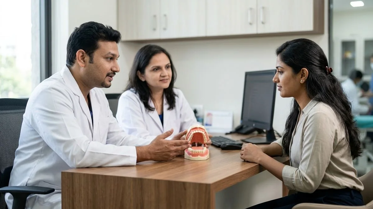
The Clinical Evaluation Process at Mouth Cancer Surgeons, Chennai
At Mouth Cancer Surgeons, we believe that no patient should have to live with the anxiety of an undiagnosed oral lesion. Our dual-surgeon team—Dr. Pradeep S. and Dr. Kalpa Pandya—provides a seamless, highly coordinated diagnostic pathway for all mucosal anomalies of the lips and oral cavity.
When you present to our clinic at Apollo Main Hospital, Greams Road, Chennai, your diagnostic journey includes:
1. Detailed Clinical History
We discuss your lifestyle habits, including tobacco use, alcohol consumption, dietary factors, and occupational sun exposure. We also document how long the spot has been present and any sensory changes you have experienced.
2. Physical Examination and Palpation
Using high-intensity lighting and magnification, we examine the lip lesion, noting its margins, color variations, and surface texture. We then gently palpate the lip to assess for deep tissue firmness (induration) and perform a thorough examination of the lymph nodes in your neck.
3. Biopsy: The Gold Standard for Diagnosis
If a white spot exhibits any red-flag features or does not resolve after conservative management, we perform a tissue biopsy. A biopsy is the only definitive way to distinguish benign hyperkeratosis or inflammation from dysplasia or early-stage cancer.
- How it Works: The procedure is performed in our outpatient clinic under local anesthesia. It is virtually painless and takes less than 15 minutes.
- Incisional/Punch Biopsy: We remove a tiny, representative sample of the white patch, along with a small margin of healthy tissue, and send it to an expert oral pathologist for histopathological analysis.
- Suturing: If necessary, we place one or two microscopic, dissolvable sutures that require no painful removal.
The Power of Our Dual-Surgeon Model
At Mouth Cancer Surgeons, we operate under a unique care delivery model. Unlike standard practices where patients are passed from one doctor to another, the same two surgeons—Dr. Pradeep S. and Dr. Kalpa Pandya—care for you from your initial diagnostic biopsy all the way through treatment and long-term follow-up.
Every biopsy report, clinical presentation, and treatment plan is reviewed by both surgeons together. This collaborative approach ensures diagnostic accuracy, minimizes patient anxiety, and delivers the highest standards of clinical safety.
Treatment and Management Pathways for Lip Lesions
The appropriate treatment for white spots on the lips is entirely dependent on the underlying cause determined by clinical examination and biopsy.
[Patient Presents with White Lip Spot]
         │
         ├──> Benign (Fordyce, Milia) ──────> Reassurance & Observation (Cosmetic removal optional)
         │
         ├──> Infectious (Thrush, HSV) ─────> Antifungal or Antiviral Therapy
         │
         └──> Suspicious (Leukoplakia, AC) ──> Diagnostic Biopsy
                                                     │
                             ┌───────────────────────┴───────────────────────┐
                             ▼                                               ▼
                     Benign Hyperkeratosis / Mild Dysplasia          Moderate/Severe Dysplasia or Cancer
                             │                                               │
                     Regular Surveillance & Risk Cessation           Wide Surgical Excision & Reconstruction
Management of Benign Lesions
- Fordyce Spots & Milia: These require no medical treatment. If they cause significant cosmetic concern, we can coordinate minimally invasive options such as CO2 laser therapy or micro-needle expression.
- Oral Thrush: Managed effectively with a course of topical antifungal oral suspensions (such as Nystatin) or systemic antifungal tablets, alongside addressing underlying causes like dry mouth or poor denture hygiene.
- Cold Sores & Canker Sores: Managed supportively with topical antiviral creams, protective oral pastes, and lifestyle modifications to reduce triggers.
Management of Precancerous Lesions (OPMDs)
If a biopsy of a white patch reveals epithelial dysplasia or actinic cheilitis, proactive intervention is necessary:
- Risk Factor Cessation: Immediate cessation of all forms of tobacco and alcohol is mandatory to stop the progression of mucosal damage.
- Surgical Excision (Lip Shave / Vermilionectomy): For extensive actinic cheilitis or dysplastic leukoplakia, we perform a precise surgical procedure to remove the damaged outer layer of the lip (the vermilion) while preserving the underlying muscle and function.
- Reconstructive Surgery: If a significant portion of the lip requires excision, our reconstructive expertise comes into play. Dr. Pradeep S. specializes in advanced reconstructive and restorative surgery, utilizing local tissue flaps to rebuild the lip, ensuring excellent cosmetic outcomes, normal speech, and complete oral continence.
Prevention and Long-Term Lip Health Strategies
Maintaining the health of your lips and oral mucosa requires consistent, proactive habits. To protect your lips from both benign irritations and serious precancerous changes, we recommend the following strategies:
- Apply Sun Protection Daily: Just like your skin, your lips need protection from ultraviolet radiation. Use a high-quality lip balm with SPF 30 or higher daily, especially if you spend significant time outdoors in sunny climates like Tamil Nadu.
- Avoid All Tobacco Products: Smoking cigarettes, bidis, or using smokeless tobacco (such as gutkha, mawa, or khaini) introduces highly carcinogenic chemicals directly to the lip tissue, serving as the primary driver of oral leukoplakia and cancer.
- Maintain Excellent Oral Hygiene: Brush and floss regularly, clean your tongue, and ensure that any dental prostheses (dentures or crowns) fit comfortably without rubbing against your lips or cheeks.
- Stay Hydrated: Drink plenty of water to keep your oral tissues hydrated, which helps maintain the protective mucosal barrier of your lips.
- Schedule Regular Oral Screenings: If you have a history of tobacco use, sun exposure, or prior oral lesions, schedule regular oral cancer screenings with a specialist. Early detection of mucosal changes significantly improves treatment success and minimizes the need for extensive surgery.
Key Takeaways
- Most white spots are harmless: Fordyce spots and milia are benign, non-cancerous, and extremely common.
- Never ignore a persistent spot: Any white spot or patch on the lip that lasts longer than two weeks, feels firm, or bleeds requires immediate professional evaluation.
- Early intervention saves lives: Catching precancerous conditions like leukoplakia or actinic cheilitis early allows for highly conservative treatments with excellent outcomes.
- Expert care matters: At Mouth Cancer Surgeons, Chennai, our dual-surgeon team provides comprehensive, compassionate care from diagnostic biopsy to advanced reconstruction.
Have questions about your condition? Request a consultation with our oral & maxillofacial surgeons — both surgeons review every case together.
For personalised treatment options and expert care, consult Dr. Pradeep S. and Dr. Kalpa Pandya — Mouth Cancer Surgeons, Chennai. Call +91 96633 03747 or book an appointment.
References
- Warnakulasuriya, S., et al. "Oral potentially malignant disorders: A consensus report from an international seminar." Journal of Oral Pathology & Medicine, 2021. [https://onlinelibrary.wiley.com/journal/13640283]
- Villa, A., et al. "Diagnosis and management of oral leukoplakia and other potentially malignant disorders." The Journal of the American Dental Association, 2019. [https://jada.ada.org/]
- World Health Organization. "Classification of Head and Neck Tumours: Epithelial Tumours of the Oral Cavity." IARC Publications, 2024. [https://publications.iarc.fr/]
- National Comprehensive Cancer Network (NCCN). "Clinical Practice Guidelines in Oncology: Head and Neck Cancers." NCCN Guidelines, 2025. [https://www.nccn.org/]
- Scully, C. "Oral and Maxillofacial Medicine: The Basis of Diagnosis and Treatment." Elsevier Health Sciences, 2013.
Authored by

Medically reviewed by