Leukoplakia: When to Worry About a White Patch in Mouth
Need expert consultation? Book an appointment with Dr. Pradeep S. or Dr. Kalpa Pandya.
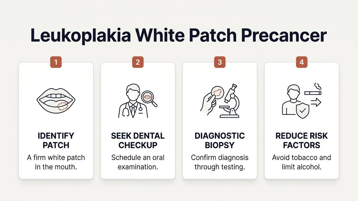
Book AppointmentDiscovering an unusual spot or texture inside your mouth can be an unsettling experience. Among the various oral lesions patients notice, a persistent, painless white patch is one of the most common reasons for referral to our practice.
In clinical terms, this unexplained white patch is often diagnosed as oral leukoplakia. Because leukoplakia is classified as an Oral Potentially Malignant Disorder (OPMD), the immediate question that arises is: leukoplakia white patch in mouth is it precancer when to worry?
Understanding the difference between a harmless tissue change and a warning sign of oral cancer is critical. In this comprehensive guide, we will break down what oral leukoplakia is, how to identify high-risk features, when you should seek immediate professional care, and how we diagnose and manage this condition at Mouth Cancer Surgeons in Chennai.
What is Oral Leukoplakia?
Oral leukoplakia is a clinical term used to describe a white patch or plaque on the mucosal lining of the mouth that cannot be rubbed or scraped off, and cannot be clinically identified as any other specific disease. It is a diagnosis of exclusion—meaning we only call it leukoplakia after ruling out other known causes of white lesions, such as oral thrush, chemical burns, or local trauma.
The World Health Organization (WHO) classifies leukoplakia as an Oral Potentially Malignant Disorder (OPMD). This classification indicates that while the patch itself is not cancer, the tissue has undergone chronic changes that make it more susceptible to transforming into oral cancer over time.
Leukoplakia can develop anywhere inside the oral cavity, but it is most frequently observed on the:
- Inner lining of the cheeks (buccal mucosa)
- Sides and under-surface of the tongue (lateral and ventral tongue)
- Floor of the mouth
- Gums (alveolar ridge)
Is Every White Patch in the Mouth Precancerous?
It is vital to understand that not every white patch in the oral cavity is precancerous or destined to become malignant. When a patient presents with a white patch, the tissue generally falls into one of several histopathological categories upon microscopic evaluation:
- Hyperkeratosis (Benign): This is a simple thickening of the outer keratin layer of the oral tissue, much like a callus on your hand. It is often a protective response to chronic irritation and carries virtually no risk of cancer.
- Mild Dysplasia: The cells show early, mild architectural and cytologic abnormalities, confined to the lower third of the epithelial layer. The risk of transformation is low, and conservative management or close monitoring may be appropriate.
- Moderate to Severe Dysplasia: The abnormal cell changes extend through the middle and upper layers of the epithelium. This is a true precancerous state. The risk of transformation into invasive oral cancer is significantly higher, requiring active intervention.
- Carcinoma in Situ: The cells are highly abnormal and resemble cancer cells, spanning the entire thickness of the epithelium, but they have not yet broken through the basement membrane into deeper tissues.
- Invasive Squamous Cell Carcinoma: Early-stage oral cancer where malignant cells have penetrated the deeper tissue layers.
Because a visual examination alone cannot determine which of these stages a white patch represents, professional assessment is essential.

Leukoplakia vs. Other White Patches: How to Tell the Difference
Many benign conditions can mimic oral leukoplakia. During your initial consultation at our Chennai clinic, we carefully differentiate leukoplakia from other common oral lesions:
| Condition | Appearance & Texture | Key Differentiating Factor | Precancerous Risk? |
|---|---|---|---|
| Oral Leukoplakia | Flat, raised, or wrinkled white patch; cannot be scraped off. | Persistent; does not resolve when local irritants are removed. | Yes (OPMD) |
| Oral Thrush (Candidiasis) | Creamy white, cottage cheese-like patches. | Can be scraped off, leaving a red, raw, occasionally bleeding surface underneath. | No |
| Frictional Keratosis | Diffuse, pale white patch, often near a sharp tooth or denture. | Resolves completely within 2–3 weeks after smoothing the sharp tooth. | No |
| Oral Lichen Planus | Lacy, web-like white lines (Wickham’s striae), often on both cheeks. | Often bilateral, symmetrical, and may cause a burning sensation with spicy food. | Very low (requires monitoring) |
| Canker Sores (Aphthous Ulcers) | Round, shallow ulcer with a white/yellow center and a bright red border. | Highly painful; heals spontaneously within 10–14 days. | No |
If you have noticed a white spot that has lasted for more than two weeks, it is important to seek an expert opinion rather than attempting to self-diagnose. If you are also experiencing persistent sores, you may want to read our detailed guide on how to identify suspicious mouth ulcers.
When to Worry: High-Risk Features of a White Patch
While all unexplained white patches deserve a professional evaluation, certain clinical features indicate a higher risk of dysplasia or malignant transformation. If you notice any of the following characteristics, you should schedule a consultation with an oral & maxillofacial surgeon without delay:
1. The Patch is "Non-Homogeneous"
Leukoplakia is broadly divided into two clinical types:
- Homogeneous Leukoplakia: The patch is uniformly white, flat, thin, and has a smooth or slightly wrinkled surface. This type has a relatively low risk of malignant transformation.
- Non-Homogeneous Leukoplakia: The patch has an uneven color or texture. It may look speckled (white mixed with red spots, known as erythroleukoplakia), nodular (bumpy), or verrucous (wart-like and thick). Non-homogeneous lesions carry a significantly higher risk of harboring severe dysplasia or cancer.
2. Location in "High-Risk" Zones
Certain areas of the mouth are highly susceptible to rapid cancer progression due to the nature of the tissue and local drainage pathways. A white patch located on the floor of the mouth, the underside or sides of the tongue, or the soft palate is considered high-risk and demands immediate attention.
3. Presence of Red Areas (Erythroleukoplakia)
If your white patch is interspersed with red, velvety areas, the risk of precancer or early cancer increases dramatically. Erythroleukoplakia has some of the highest malignant transformation rates among all oral precancers.
4. Associated Symptoms
While early leukoplakia is typically painless, you should worry if the patch begins to:
- Bleed easily when touched or brushed
- Develop a firm, hardened base (induration)
- Cause difficulty in chewing, swallowing, or moving your tongue
- Cause a persistent, localized burning sensation or pain
Common Causes and Risk Factors
Oral leukoplakia is primarily a reaction to chronic mucosal irritation. In South India, and Chennai specifically, we see a distinct pattern of risk factors driving these tissue changes:
- Tobacco Use: Smoking cigarettes, bidis, or cigars, and using smokeless tobacco (gutka, mawa, khaini) are the leading causes. The heat and toxic chemicals cause cellular damage and protective thickening of the mucosa.
- Areca Nut (Paan) Chewing: Chewing betel quid or areca nut formulations is highly prevalent. This habit is directly linked to both leukoplakia and oral submucous fibrosis (OSMF).
- Alcohol Consumption: Alcohol acts as a solvent, making the delicate oral mucosa more permeable to tobacco-associated carcinogens.
- Chronic Mechanical Trauma: A sharp, broken tooth, an ill-fitting dental crown, or a poorly designed denture that constantly rubs against the cheek or tongue can cause localized white patches (frictional keratosis) which, if left unaddressed for years, can undergo dysplastic changes.
- Idiopathic Leukoplakia: In some cases, patients develop leukoplakia without any identifiable risk factors. Interestingly, idiopathic leukoplakia (especially in non-smoking women) often carries a higher risk of malignant transformation.
The Role of a Biopsy in Diagnosing Oral Leukoplakia
You cannot determine if a white patch is precancerous simply by looking at it. A tissue biopsy is the definitive gold standard for diagnosing oral leukoplakia and evaluating the presence and severity of dysplasia.
What Happens During an Oral Biopsy?
Many patients are anxious about the word "biopsy," but the procedure is straightforward, safe, and virtually painless:
- Local Anesthesia: We administer a small amount of local anesthetic to completely numb the area around the white patch.
- Sample Collection: Using a specialized punch tool or a scalpel, we gently remove a tiny, representative piece of the white tissue (and a small edge of normal tissue for comparison).
- Suturing (if needed): If required, one or two dissolvable stitches are placed to close the site.
- Pathology Lab Analysis: The tissue sample is sent to a specialized oral pathologist who examines the cellular architecture under a microscope to check for dysplasia or early cancer cells.
At Mouth Cancer Surgeons, we perform these diagnostic biopsies in our comfortable outpatient clinic setting, ensuring patient comfort and rapid turnaround times for pathology reports.
Treatment Options and Management Strategies
If you are diagnosed with oral leukoplakia, your treatment plan will depend on the biopsy results, the size of the lesion, and its location. Our primary goal is to prevent the patch from transforming into oral cancer.
To learn more about our comprehensive approach to managing these conditions, visit our dedicated specialty page on oral precancer (OPMD) care.
1. Elimination of Irritants and Lifestyle Modification
The first step for any white patch is removing the underlying cause. This includes:
- Complete cessation of tobacco, betel nut, and alcohol.
- Dental restoration to smooth down sharp teeth, repair broken fillings, or adjust ill-fitting dentures.
- In some cases of simple hyperkeratosis, removing the irritant is enough to make the white patch disappear completely over several weeks.
2. Surgical Excision or Laser Ablation
If the biopsy reveals moderate to severe dysplasia, we highly recommend removing the lesion. This can be accomplished via:
- Cold Scalpel Excision: Complete surgical removal of the patch with a safe margin of healthy tissue. This is highly effective and allows the pathologist to examine the entire removed specimen.
- Laser Surgery (CO2 Laser): Using a precise laser beam to vaporize or excise the abnormal tissue. Laser surgery minimizes bleeding, reduces post-operative pain, and promotes rapid healing with minimal scarring.
3. Long-Term Surveillance
Even after complete surgical removal, leukoplakia has a recurrence rate of up to 10% to 20%. Therefore, long-term follow-up is mandatory. We monitor our patients every 3 to 6 months to catch any early signs of recurrence or new lesions immediately.
To understand the broader spectrum of early mucosal changes, you may also find our guide on oral cancer screening helpful.
The Dual-Surgeon Advantage at Mouth Cancer Surgeons, Chennai
When dealing with a potentially precancerous condition like oral leukoplakia, having highly specialized, experienced surgeons in your corner makes all the difference.
Mouth Cancer Surgeons is a dedicated oral & maxillofacial oncology practice in Chennai led by the dual-surgeon team of Dr. Pradeep S. and Dr. Kalpa Pandya.
Unlike larger, generalized hospital departments where you might see a different doctor at every visit, our practice operates on a unique, personalized care model:
- Consistent Dual-Surgeon Care: Both Dr. Pradeep and Dr. Kalpa personally review, diagnose, and manage every single case. The same two surgeons who perform your initial clinical evaluation will perform your biopsy, guide your treatment, and conduct your long-term follow-up surveillance.
- Specialized Expertise: Dr. Pradeep S. (MDS, FHNO, FIBCSOMS) is an internationally board-certified head & neck surgical oncologist with over 7 years of specialized experience in oral cancer and reconstructive surgery. Dr. Kalpa Pandya (MDS, FHNS) brings over 10 years of experience and has successfully managed more than 1,000 oral cancer and precancer patients, leading our OPMD and rehabilitation services.
- World-Class Infrastructure: We consult and operate primarily at Apollo Main Hospital, Greams Road, Chennai, providing our patients access to cutting-edge diagnostic tools, advanced laser technologies, and a multidisciplinary team of oncologists if required.
If you are experiencing a persistent white patch or have concerns about oral precancer, early intervention is key. Book a comprehensive consultation with Dr. Pradeep S. and Dr. Kalpa Pandya at Apollo Main Hospital, Greams Road, Chennai.
For personalized treatment options and expert care, consult Dr. Pradeep S. and Dr. Kalpa Pandya — Mouth Cancer Surgeons, Chennai. Call +91 96633 03747 or book an appointment.
References
- Warnakulasuriya, Saman, et al. "Oral potentially malignant disorders: A consensus report from an international seminar." Oral Diseases, 2020. https://onlinelibrary.wiley.com/doi/10.1111/odi.13375
- World Health Organization. "Classification of Tumours: Head and Neck Tumours." IARC Press, 2017.
- Villa, Alessandro, et al. "Diagnosis and management of oral leukoplakia: A systematic review." Journal of the American Dental Association, 2017. https://jada.ada.org/article/S0002-8177(16)30908-1/fulltext
- National Comprehensive Cancer Network (NCCN). "Clinical Practice Guidelines in Oncology: Head and Neck Cancers." NCCN, 2024. https://www.nccn.org/guidelines
- Speight, Paul M., et al. "The natural history and malignant transformation of oral epithelial dysplasia." Oral Surgery, Oral Medicine, Oral Pathology, Oral Radiology, 2018.
Authored by

Medically reviewed by